Cancer cell migration pack
Follow long-term cancer cell migration in real time on top of the microscope
Keep ideal temperature conditions
Maintain your cells on top of the microscope for longterm assays
Follow cancer cell migration in ral time
Improved quality of image data gathering
Adapted to cultures under flow
Designed to fit different tubing diameters

Need a microfluidic SME partner for your Horizon Europe project?
Cancer cell migration pack
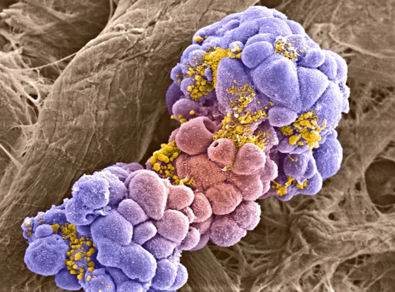
Cancer cell migration is a leading factor in poor patient prognosis
Cancer cell migration, also known as metastasis, is the process by which cancer cells detach from the original tumor to colonize another body area.
It is one of the leading causes of cancer mortality, accounting for 90% of the cases and reducing the 5-year survival rate to less than 30% [1].
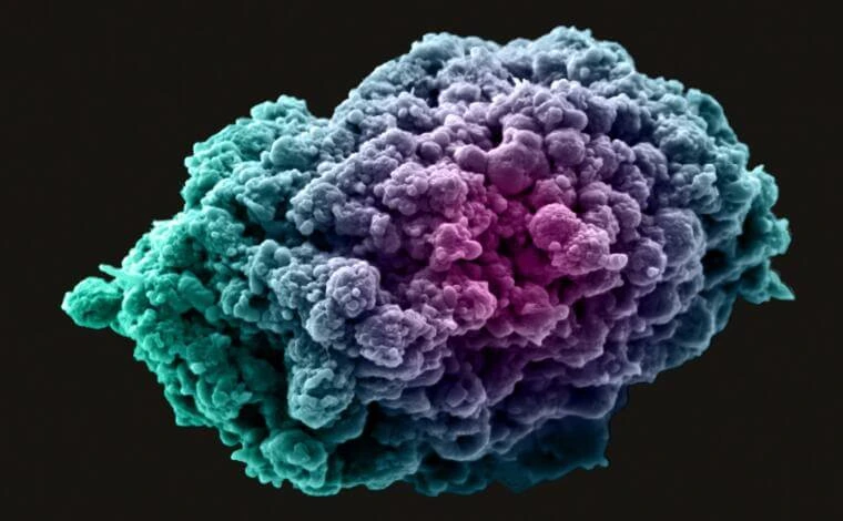
Several factors can affect cancer cell migration
The tumor microenvironment plays a role in the migration of metastatic cells. As the tumor grows, the interior of the cancer mass starts to receive fewer nutrients and oxygen.
The starvation and, especially, the hypoxia lead to changes in the secreting pattern of the cells, which can affect the motility and even the drug resistance of the tumor [2].

The mechanisms for cancer cell migration are still not precise
Tumors are famous for their internal heterogeneity, so bulk research overlooks potentially essential differences between cell types that could indicate ways of preventing metastasis.
New live cell imaging techniques have unraveled some of these mysteries, but they require cells to stay on top of the microscope stage for long periods, challenging ideal cell culture conditions.
References
1. Wang, Y; Wu, D; Wu, G; Wu, J; Lu, S; Lo, J; He, Y; Zhao, C; Zhao, X; Zhang, H; Wang, S. “Metastasis-on-a-chip mimicking the progression of kidney cancer in the liver for predicting treatment efficacy”. Theranostics. 2020 Jan 1;10(1):300-311. doi: 10.7150/thno.38736. PMID: 31903121; PMCID: PMC6929630.
2. Nam, H; Funamoto, K; and Jessie, J. “Cancer cell migration and cancer drug screening in oxygen tension gradient chip”, Biomicrofluidics 14, 044107 (2020) doi.org/10.1063/5.0011216
3. Huang, Y; Agrawal, B; Clark, P. A; Williams, J. C; Kuo, J. S. “Evaluation of Cancer Stem Cell Migration Using Compartmentalizing Microfluidic Devices and Live Cell Imaging”. J. Vis. Exp. (58), e3297, doi:10.3791/3297 (2011).
Improved imaging for cancer cell migration
Temperature is one of the critical parameters that need to be kept constant during long-term live cell imaging. Our stage-top incubator was designed to do precisely that, especially for dynamic cell culture and underflow experiments. Also, the stage-top incubator ensures that the flowing liquid arrives at the cells at the desired temperature.
The standard setup is displayed below. It can easily be customized depending on the application.

The cancer cell migration pack includes:
Flow sensor (Galileo, MIC)
Stage top incubator (in development)
Software (Galileo user interface)
Flow controller
2 x 15 mL falcon reservoirs
Chip from microfluidic ChipShop (suggestion: Fluidic 834)
All necessary accessories: tubing, connectors, filters, etc.
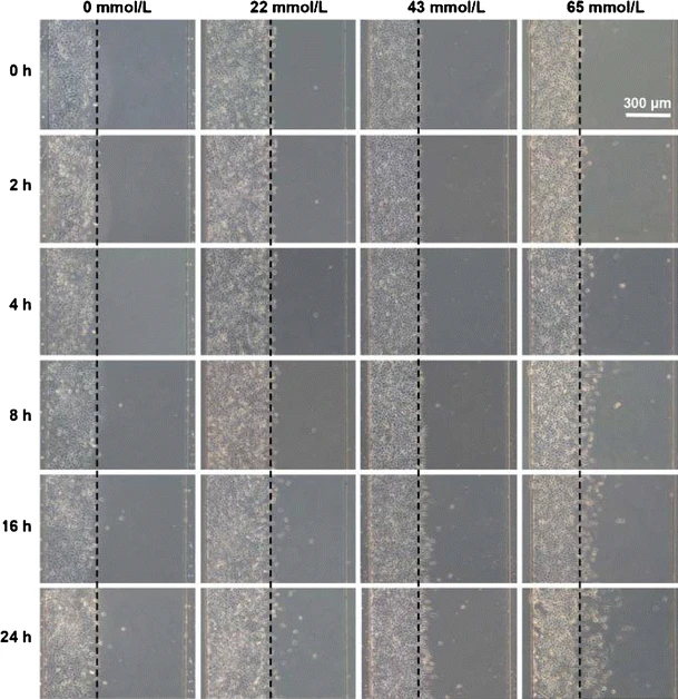
The effect of Ethanol on cancer cell migration
Huang et al. [1] studied breast cancer cell migration under the influence of ethanol. Among other factors, such as smoking, ethanol consumption has been reported to be an independent risk for breast cancer development and progression [2], as it can affect estrogen levels in the blood [3].
They cultured breast cancer cells under flow on a chip. Once confluent, they simulated a wound using laminar flow to apply trypsin to only a part of the chip. Then, they observed the cells while exposing them to different ethanol concentrations.
Over 24h, the highest ethanol concentration (65 mmol/L) induced the highest cell migration of the tested conditions, which showed an overall direct correlation. Studies such as this pave the way for scientists to study cells under a controlled microenvironment so the nuances of cellular behavior can be clarified.
References
1. Huang, X., Li, L., Tu, Q. et al. On-chip cell migration assay for quantifying the effect of ethanol on MCF-7 human breast cancer cells. Microfluid Nanofluid 10, 1333–1341 (2011). https://doi.org/10.1007/s10404-011-0766-9
2. Boffetta P, Hashibe M (2006) Alcohol and cancer. Lancet Oncol 7:149–156
3. Maniyar R, Chakraborty S, Suriano R. Ethanol Enhances Estrogen Mediated Angiogenesis in Breast Cancer. J Cancer. 2018 Oct 6;9(21):3874-3885. doi: 10.7150/jca.25581. PMID: 30410590; PMCID: PMC6218769.
Customize your pack
Contact our experts to answer any questions about this stage top incubator for microfluidics pack and how it can match your specifications!
Frequently asked questions
Can the stage top incubator be sterilized?
Yes, we have developed a simple protocol for sterilization and cleaning that is provided along with the user guide.
Can the stage top incubator be placed inside the CO2 incubator?
The Stage Top Incubator was designed to be kept outside the CO2 incubator, but it can be adapted on demand.
Is the stage top incubator gas-tight?
No, the Stage Top Incubator allows gas exchange with the atmosphere.
Do the reservoirs of media need to be kept at 370°C?
No, the reservoirs can be kept at room temperature. The Stage Top Incubator was designed to ensure that the media reaches the cells at the desired temperature regardless of the temperature of the reservoirs.


Funding and Support
Products & Associated Accessories
FAQ - Cancer Cell Migration
What exactly is the “Cancer Cell Migration Pack”?
It is a microfluidic system designed to allow real-time monitoring of cancer cell movement on a microscope over a long period of time. The point is easy enough, keep the cells in controlled conditions (temperature in particular) and you image them over hours, even a day, without the typical tradeoffs that accompany the long-term presence of cells on the microscope stage.
Why cell migration in cancer cells is such a big issue to researchers working on metastasis?
Since migration is a characteristic stage of metastasis, cells dissociate from a primary tumor and migrate to a different location. Clinically, it is generally accepted that metastasis is the primary motive of cancer deaths; this page refers to the numbers commonly used in the context (approximately 90% of cancer deaths are metastatic and 5-year survival in this scenario is less than 30%). Migration is difficult to overlook, whether your discipline is the basic mechanisms or the translational screening.
What is the added value in microfluidics to the “classic” migration assays (scratch assay, Transwell, etc.)?
Classic assays are convenient, but distort reality: they are in more realistic statistics, and they have less gradient control and average out heterogeneous behaviors. Microfluidic migration models provide this ability to tune flow, nutrient delivery and microenvironmental constraints with much further precision, and are compatible with continuous imaging. It is of particular importance when you want to measure specific or subpopulation-specific behaviors instead of a bulk readout.
What is in the standard configuration?
A typical installation that is customizable can be described in the page, although the minimum setup includes:
- A flow sensor (Galileo, MIC)
- A stage-top incubator (referred to as in development)
- User interface control software (Galileo user interface)
- A flow controller
- Two 15 mL Falcon reservoirs
- An example microfluidic ChipShop (currently called Fluidic 834) microfluidic chip proposal.
- Accessories like tubing, connectors, filters, etc. with consideration of compatibility between various tubing sizes.
Is it possible to migrate assays in flow without destroying my image?
That is one of the points of the pack. It is placed as adapted to cultures under flow with better quality of image data collection, namely due to the fact that the platform is expected to maintain cells in long term viability and stability on the microscope even as the fluid is perfused. Practically this is where attention to thermal control and the measurement of steady flow begin to become valuable than supposed.
Does it assist in tumor microenvironment such as hypoxia or nutrient deprivation?
Yes, indirectly, as it provides the ability to control microenvironment and observe it over a long period. It emphasizes that the tumor microenvironmental limitations (depleting nutrients and oxygen) can alter the pattern of secretion, motility and drug resistance and that microfluidic manipulation and live imaging is a viable path forward to investigate such effects wherein fewer black box variables exist in the presence of microfluidic control compared to mono-culture. In case your work particularly requires oxygen tension gradients, you would normally add that as an overlay control layer to the expanded platform design.
Can you give an example of a concrete experiment this pack is intended for?
An on-chip migration experiment of the ethanol impact on breast cancer cells (MCF-7). In that case, cells were cultured in a flowing condition; a simulation of a wound was done through laminar flow to localize the exposure of trypsin; and migration was followed at various concentrations of ethanol. The most tested condition of ethanol (65 mmol/L) yielded the greatest migration of the conditions tested over 24 hours indicating a dose-linked effect in that arrangement.
Is it possible to sterilize the stage-top incubator, and how painful is cleaning?
Yes, it is, according to the page: there is a straightforward sterilization and cleaning procedure that is supplied with the user manual. This counts in real laboratory life, since sterilizable is a relative term, the other half is whether it gets done consistently.
Should I place the stage-top incubator in a CO2 incubator to ensure that the cells are happy?
Not by default. The stage-top incubator is programmed to be positioned outside of the CO2 incubator (to maintain your microscope work process simple), but can be customized on the fly. Also important to note: it is said to be not gas-tight i.e. allow gas exchange with the ambient atmosphere.
Are media reservoirs to be maintained at 37 °C temperatures in the course of perfusion?
No. The page even states that reservoirs may be kept at room temperature, since the stage-top incubator is designed such that the medium would be at the desired temperature at the cells despite the temperature of the reservoir. Quite a convenient detail–considering that long-run operation with hot reservoirs may be active and has additional failure modes.
We are creating a Horizon Europe consortium: why do we need a microfluidics SME partner (such as MIC) in this case?
Should you require microfluidic platform engineering, microfluidic platform prototyping, microfluidic platform integration with imaging, or with powerful experimental setups that work on Day One, an SME that already designs and manufactures such systems can de-risk the technical work packages. And, in reality, when implementation, prototyping, and exploitation plans are in the hands of an expert SME, they tend to increase the plausibility of the science, which is what evaluators examine when the science is high but the path to validation remains vague.
