Microfluidic Flow Cytometry Instrument Pack
Hydrodynamic sheath focusing for microfluidic flow cytometer systems
Microfluidic pack for cytometry
Unbox, set up and start your experiments right away
Pulsation free flow controller
Stable flow focusing with a flow controller
No cell aggregation
Cell focusing in a narrow stream inside a wide channel

Need a microfluidic SME partner for your Horizon Europe project?
Drawing adapted from Neil Dufton (wellcome collection CC BY 4.0); Sheath flow principle drawing by Torino et al. (2017)
Microfluidic flow cytometry
Sheath flow focusing cytometry in a microfluidic channel is now available using the microfluidic flow cytometry Pack. This method detects and measures cells’ relevant chemical and physical characteristics, with the possibility of sorting them. For example, cytometry can be used to discriminate if a cell died from necrosis or apoptosis or to perform cell proliferation assays.
Based on a fast and accurate flow controller and sheath flow chip, this all-in-one solution contains all the required microfluidic pieces for researchers to set up their microfluidics cytometry system with excellent sensitivity. Moreover, our microfluidic specialists will help you set up your platform with beginner-friendly advice and user guides.
Sheath flow streams aim to align the cells in the center of the microfluidic channel for cytometry. A standard sheath fluid flow focusing cytometry pack contains two pumping channels: the first pushes the sample to the microfluidic chip, and the second pumps the sheath fluid. The stability of focusing is directly linked to the flow rates of the sample stream and the sheath streams.
Thanks to our wide-range flow sensor (Galileo), flow rates can be measured and regulated. For more info about the Galileo project, follow this link.
The microchannel material (either PMMA or COP) is optically transparent, with low dispersion and birefringence, which makes it suitable for cytometry. Therefore, a light detection system for cytometry can be set up directly on top of the microfluidic channel.
Cell sorting is possible thanks to the five outlet channels with two junctions, which allow the collection of target cells at two different locations by applying positive or negative pressure at the outlet.
Flow cytometry pack setup
An all-in-one pack guarantees good compatibility between the different instruments, allows you to start your experiment immediately, is piloted by a single software, and can be used for other applications. These are a few arguments why a pack is the easiest way to setup a microfluidic experiment.
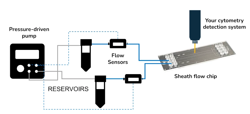
A typical flow cytometry pack contains:
Flow sensor (Galileo, MIC)
Software (Galileo user interface)
Flow controller
Three reservoirs
Microfluidic chip from microfluidic ChipShop
All necessary accessories: connectors, tubing, filters, etc…
Microfluidic flow cytometry principle
Microfluidic flow cytometers have better portability and disposability than conventional devices, in addition to reduced sizes and costs [1]. This system is used to investigate single-cell level biological events and can also be used for cell sorting [2].
Microfluidic channels with laminar flows allow better accuracy and more stable flow control than conventional cytometry techniques like glass capillary-based systems [3]. The sheath fluid can also be air in a hydrophobic microfluidic channel (made of either PMMA or COP) to create a disposable air sheath flow cytometry system [4].
Brillouin flow cytometry spectroscopy has been performed in a microfluidic channel to describe the mechanical properties of the cells [5].
Finally, integrated microfluidic cytometry has also been adapted for on-point measurements [6].
Advantages of microfluidics for flow cytometry
Conventional flow cytometry consumes large sample and reagent volume, making the experiment expensive. Moreover, classical flow cytometry systems are mechanically complex, requiring trained personnel for maintenance, and are usually complicated to customize for specific applications.
Microfluidics allows the fabrication of miniaturized flow cytometers that require less sample and reagent volume, which reduce the cost of the experiment and are suitable for implementation in remote environments. The microfluidic sheath flow cytometry system has an exceptional sensitivity for single-cell analysis as various detectors can be used to experiment.
Moreover, fluid properties can be precisely tuned at the microfluidic scale, and very stable focused sample flow can be achieved thanks to a strictly controlled flow rate on both the sample and the sheath streams.
Sheath flow cytometry can also be coupled with cell sorting directly in the same chip.
Microfluidic sheath flow cytometry allows more integrated, precise, and cheaper cell analysis.

References
TD Chung, HC Kim, Recent advances in miniaturized microfluidic flow cytometry for clinical use, Electrophoresis 2007, 28, 4511–4520
Fu, A. Y., Chou, H. P., Spence, C., Arnold, F. H., Quake, S. R., An integrated microfabricated cell sorter Anal. Chem. 2002, 74, 2451–2457.
Huh, D., Gu, W., Kamotani, Y., Grotberg, J. B., Takayama, S., Microfluidics for flow cytometric analysis of cells and particles Physiol. Meas. 2005,26, R73–R98.
Huh D, Tung Y-C, Wei H-H, Grotberg J B, Skerlos S J, Kurabayashi K and Takayama S 2002 Use of air-liquid two-phase flow in hydrophobic microfluidic channels for disposable flow cytometers Biomed. Microdevices 4141–9
Zhang J et al., Brillouin flow cytometry for label-free mechanical phenotyping of the nucleus, Lab Chip, 2017, 17, 663
Kiichi S., Akihide H., Mnabu T., Hideaki H., Takehiko K.; Microchip-based chemical and biochemical analysis systems, Advanced Drug Delivery Reviews, 55, 3, 24 February 2003, Pages 379-391
Customize your microfluidic flow cytometry pack
To achieve good cell separation and analysis, the sheath stream flow rate should be much higher than the sample stream flow.
The sheath flow cell sorting chip is available in poly(methyl methacrylate) (PMMA) or Cyclo Olefin Copolymer (COP) materials. While both are suitable for cytometry, COP has better optical properties, and PMMA is slightly stronger.
There are two different chips, each with two channels available. The first one has a 50 µm depth structure in its first channel and a 25 µm depth in its second. The second one has both 30 µm depth channels.
If necessary, the pack can be equipped with additional pumping channels for the OB1 flow control pump and flow rate sensors.
Bubbles can be a problem for cells and cytometry; if necessary, a bubble remover that tackles this issue can be provided.
Frequently asked questions
How can we help your experiment?
This pack is in beta testing phase. So, although the instruments are not fully industrialized, we can provide extensive support as part of our beta testing program. Get in touch to see if you are eligible.
Can a pack be customized based on my specific application?
Yes! Our experts will establish which instruments are best suited for your application, such as the type of flow sensor or the number of flow controller channels you need to perform your experiment. Contact us using the “talk to our experts” green button above.
Can I buy individual packs?
Our instruments are in beta testing phase and can be tested as a pack or individually, so get in contact with our team to know how our beta testing program works.
Download the MIC Horizon Europe 2026/2027 Calls Calendar:
Products & Associated Accessories
FAQ - Microfluidic Flow Cytometry Pack
What is the Microfluidic Flow Cytometry Pack?
The Microfluidic Flow Cytometry Pack is a system of instruments in one that enables sheath-flow focusing cytometry within a microfluidic channel. It identifies and quantifies chemical and physical properties of single cells and has the added advantage of sorting them. Typical applications include distinguishing between apoptotic and necrotic cell death, conducting cell proliferation assays, and performing biological analysis at the single-cell level in a customizable, small-scale format.
What is the operation of the sheath flow focusing in this system?
The cells are centered within the microfluidic channel using sheath-flow streams. These two pumping channels confine the cells into a small, focused stream within a larger channel by carefully regulating the relative flow rates of both streams to the cells and the sheath fluid. This single-file alignment ensures that each cell passes through the detection zone individually, which is important for accurate measurement and to avoid cell aggregation.
What are the main strengths of using microfluidic flow cytometry compared to conventional systems?
Traditional flow cytometry also requires large sample and reagent volumes, making experiments costly. Mechanically complex systems, such as classical systems, also need trained staff to maintain their functioning, and are hard to tailor to a particular purpose. Microfluidic flow cytometry overcomes these disadvantages by making the system smaller, reducing the amount of sample and reagents used, keeping costs down, and enabling easier integration into remote settings. The laminar flow properties of microchannels also give the system greater control over flow, yielding more stable and precise results than glass capillary-based methods.
Is it possible to use this pack in cell sorting and cell analysis?
Yes. The five outlet channels with two junctions prevent the cell sorting process because the chip used in the analysis process can be used to collect the targeted cells at two points with either positive or negative pressure applied at the outlet, an important benefit of the integrated low-volume workflow.
What are the available chip materials, and what are some of their distinctions?
The sheath flow cell sorting chip is offered with poly(methyl methacrylate) (PMMA) or Cyclo Olefin Copolymer (COP). Both can be used in cytometry; however, COP has superior optical characteristics, with low dispersion and birefringence, while PMMA is slightly more mechanically robust. Both materials are optically transparent with low dispersion and birefringence, making them well-suited for mounting a light detector system over the microchannel. They are interchangeable based on the higher priority of the given application, with either optical clarity or mechanical durability selected.
How many channel dimensions are in the chips?
Two chip formats are offered. The first channel of the first has a 50-um depth structure, and the second has a 25-um depth. Each channel of the second chip is 30 μm deep. These dimensions are set to follow the cells through the detection region of a single focused stream, and the chip geometry must be chosen to match the dimensions of the cell or particle size range under analysis.
What are the instruments of a normal pack?
A standard flow cytometry package includes the flow controller, Galileo flow sensor, three reservoirs, a microfluidic chip from the microfluidic Chipshop, all accessories, including connectors, tubing, filters, and the Galileo user interface software.
What is the method of flow stability during cytometry experiments?
This is the key to good cytometry results, and the pack addresses this by using its pulsation-free pressure-driven flow controller. Since the quality of cell focusing is directly proportional to the sample-to-sheath flow-rate ratio, real-time monitoring and control enable the narrow, stable stream essential for fine single-cell detection. To maintain a constant focus, the sheath stream flow rate is kept significantly higher than the sample stream flow rate.
Are bubbles capable of messing up the experiments, and can this be avoided?
Yes, cytometry experiments may be affected by air bubbles during cell viability and optical detection. If a specific experiment requires concern about bubbles, a bubble remover can be attached to the pack to resolve the issue. This is an optional accessory and can be included in the customization process when discussing instrument needs with our team.
Is the pack on sale in the market, and is it customizable?
The pack is under beta testing. The instruments are not yet fully industrialized, but they are extensively supported under the beta testing program. Customization is free of charge; our team may advise on the optimal type of flow sensor, the number of flow controller channels, the chip material, and other accessories, depending on the type of experimental work. Recent studies that seek to test the pack separately or in the whole system can contact our team directly to determine eligibility and needs for the pack.
