DNA-Paint Pack
Recognize and bind DNA sequences specifically
Study the location and organization of specific genomic regions
Probe many targets simultaneously
Use different samples: fixed cells, live cells, or tissues
Single-molecule localization microscopy
Single-molecule localization microscopy (SMLM) is an effective tool to investigate small structures of biological systems thanks to its unprecedented resolution. At the forefront of these developments, there is DNA-paint (DNA-based point accumulation for imaging in nanoscale topography).
It is a rapidly developing fluorescence super-resolution technique that exploits the stochastic and transient binding of fluorescently labeled DNA probes. In its early stages, the implementation of DNA-PAINT was limited by its low throughput, excessive acquisition time, and difficult integration with live-cell imaging [1].
DNA-paint has multiple advantages such as reaching spatial resolutions below 10 nm and enabling the imaging of multiple targets in the same sample [2].
We offer you a versatile computer-controlled microfluidic setup to enable multiplexed DNA-PAINT in an efficient manner.
DNA-paint pack principle
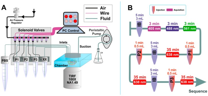
The technique uses base pairing between fluorescently labeled short DNA oligonucleotides [3]. The target is tagged with a short DNA docking strand, while the complementary fluorescently labeled imager strand freely diffuses in solution. Upon hybridization, an increase in fluorescence intensity is observed for several hundred milliseconds (ON), after which the imager strand is detached (OFF), leaving the docking strand empty. As the imager strands are bound and unbound, the pool of imaged fluorophores is continuously replenished, eliminating the photon budget limitation in DNA-PAINT [4].
Advances have reduced the limiting factors that determine the binding frequency, speeded up image acquisition, and enabled DNA-PAINT imaging in minutes. The complex environment of living cells poses many challenges to standard DNA-based point accumulation for imaging in nanoscale topography imaging. These challenges are addressed by improved probe designs, like using modified DNA nucleotide or amino acid-based backbones for imager and docking probes [4].
DNA-paint pack setup
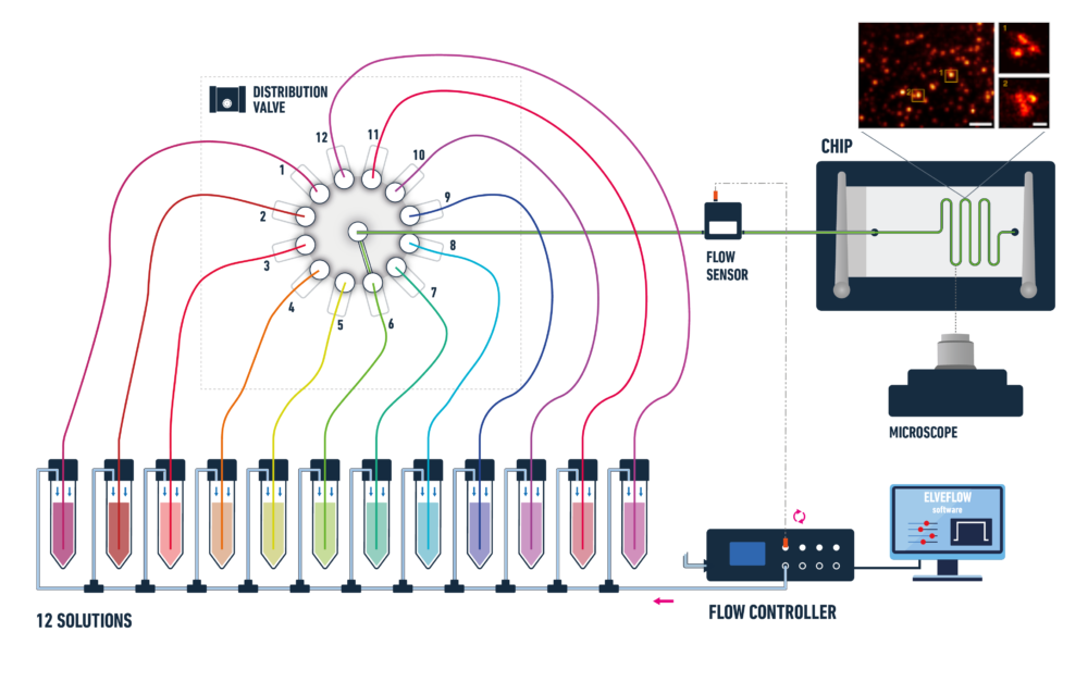
This is an illustrative setup example. The solutions’ number and volume can be adjusted according to the experimental requirements and protocol.
Pseudodiffraction-limited fluorescence image of DNA origami labeled with DNA-PAINT docking strands by Deußner-Helfmann et al., 2018.
A typical pack contains:
- OB1 flow controller (Elveflow)
- A flow sensor (MFS or BFS) (Elveflow)
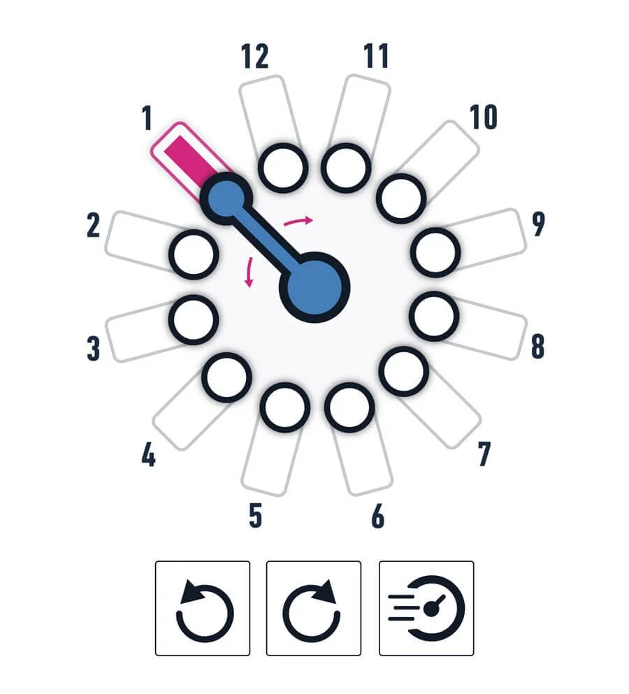
- One or two 12:1 MUX distribution valves (Elveflow) depending on the number of dyes you want to inject
- Tubings and connectors
- Several Eppendorfs or Falcon reservoirs
- Microfluidic chips* advice and recommendation
- Automation and control software (Elveflow)
- A user guide
The solution can be configured as per user’s requirements. Our experts can help you define the best possible setup to reach your goal.
Thanks to the versatility of the instruments, and the upgradability of our range, the pack can be adapted to your needs.
Advantages
Thanks to the high performance of the flow controller, you will be able to:
- Have ultra-precise flow control of small dispensed volumes.
- Synchronize with other equipment, such as a fluorescence microscope.
- Improve reproducibility.
- Have a fast and easy sequential injection system for different solutions.
- Automate your experiments*.
- Have simultaneous visualization of multiple genomic regions.
- Have high specificity of DNA sequences.
- Have a high spatial resolution.
*The sequence scheduler automatizes the platform to flow a large number of different solutions easily. Our experts can help you integrate your experiment using TTL triggers or direct software integration via SDK.
DNA-paint pack proof-of-concept
As proof of principle, labeled and imaged proteins on mitochondria, the Golgi apparatus, and chromatin were used in the paper “Nanobody Detection of Standard Fluorescent Proteins Enables Multi-Target DNA-PAINT with High Resolution and Minimal Displacement Errors” by Shama Sograte-Idrissi et al. [2].
References
1. van Wee, R., Filius, M., & Joo, C. (2021). Completing the canvas: advances and challenges for DNA-PAINT super-resolution imaging. Trends in Biochemical Sciences, 46(11), 918-930.
2. Sograte-Idrissi, S., Oleksiievets, N., Isbaner, S., Eggert-Martinez, M., Enderlein, J., Tsukanov, R., & Opazo, F. (2019). Nanobody detection of standard fluorescent proteins enables multi-target DNA-PAINT with high resolution and minimal displacement errors. Cells, 8(1), 48.
3. Jungmann, R., Steinhauer, C., Scheible, M., Kuzyk, A., Tinnefeld, P., & Simmel, F. C. (2010). Single-molecule kinetics and super-resolution microscopy by fluorescence imaging of transient binding on DNA origami. Nano letters, 10(11), 4756-4761.
4. van Wee, R., Filius, M., & Joo, C. (2021). Completing the canvas: advances and challenges for DNA-PAINT super-resolution imaging. Trends in Biochemical Sciences, 46(11), 918-930.
DNA-paint pack applications
DNA-based point accumulation for imaging in nanoscale topography (DNA-paint) has many applications in biological research, including the study of genome organization, gene expression, and chromatin structure. It allows researchers to visualize the location of specific DNA sequences within cells and tissues, providing insights into the structure and function of the genome.
Here are some examples:
- Genome organization: The technique can be used to study the organization of the genome within the nucleus. By labeling specific DNA sequences with fluorescent probes, researchers can visualize the location of these sequences within the nucleus and study their spatial relationships with other genomic regions.
- Gene expression: DNA-paint can be used to study gene expression patterns. Labeling the RNA transcripts of specific genes with fluorescent probes makes it possible to track the location and dynamics of these transcripts within the cell.
- Chromatin structure: The technique can be used to study the structure of chromatin, the complex of DNA, and proteins that make up chromosomes. By labeling specific chromatin regions with fluorescent probes, their organization and dynamics within the nucleus can be studied.
- Diagnosis and disease research: DNA-paint can be used in diagnostic assays and disease research. By designing probes that recognize specific disease-associated DNA sequences, the technique is used to detect and study disease-related changes in the genome.
- Nanotechnology: such as DNA-based nanodevices and biosensors. Using DNA as a scaffold, researchers can build complex structures and devices with precise control over their size and shape.
How can we help your experiment
The packs applications are still under development, so we are not able to give you the tips or troubleshooting advice that we usually give in our user guides and application notes for the possible challenges you could face for a specific application experiment. That being said, we can always guarantee reliable and high-precision microfluidic flow control. Our microfluidic instruments are high-performance, versatile and user-friendly. Our experts will bring support and expertise during the setup and implementation of this sensor calibration pack.
Can a pack be customized
Yes! Our experts will establish which instruments are best suited for your application, such as the type of flow sensor or the number of flow controller channels you need to perform your experiment. Send us a message at: innovation@microfluidic.fr
Can I buy individual instruments?
You can order our instruments on the product section of our website.