Liver-on-a-chip model
Mimic the liver microenvironment in long term experiments
Relevant microenvironment
Improve your reproducibility with physiological culturing conditions
Up to 3-weeks long cultures
Automated and controlled supply of nutrients in a stable flow
Up to 4 parallel cultures
Test different conditions at the same time

Need a microfluidic SME partner for your Horizon Europe project?
Image credit: High-speed BCARS allows detailed mapping of specific components of tissue samples. A false-color BCARS image of mouse liver tissue (left) picks out cell nuclei in blue, collagen in orange and proteins in green. An image of tumor and normal brain tissue from a mouse (right) has been colored to show cell nuclei in blue, lipids in red and red blood cells in green. Images show an area about 200 micrometers across. Credit: Camp/NIST.
Why use liver-on-a-chip models?
The quest for more reproducible models to advance drug development and disease research is in full swing. Current animal models are known to have limitations when translating from preclinical to human trials, especially when considering effects on the liver.
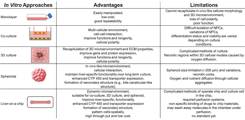
A striking example is that almost half of the drugs found safe in established animal models were responsible for liver damage in clinical trials [1]. Thus, there are several approaches to improve translation between preclinical and clinical data, as illustrated in the table below [adapted from 1].
Every method has shortcomings, but liver-on-a-chip models offer the possibility to perform all the in vitro models, taking benefits from their advantages. As a new technology, there are no standards yet, and operating organ-on-a-chip systems requires specialized know-how. This is precisely what we are aiming to simplify with our platform.
Advantages and limitations of in-vitro liver models [1]

We have recently published a review about the different organ-on-a-chip models and current innovations.
References
1. Deng J, Wei W, Chen Z, Lin B, Zhao X. Engineered liver-on-a-chip platform to mimic liver functions and its biomedical applications: a review, Micromachines. 2019; 10(10):676. https://doi.org/10.3390/mi10100676
2. Shin JY, Park J, Jang HK, Lee TJ, La WG, Bhang SH, Kwon IK, Kwon OH, & Kim BS. Efficient formation of cell spheroids using polymer nanofibers. Biotechnology letters. 2012; 34(5):795–803. https://doi.org/10.1007/s10529-011-0836-9
3. Moriyama M, Sahara S, Zaiki K, Ueno A, Nakaoji K, Hamada K, Ozawa T, Tsuruta D, Hayakawa T, & Moriyama H. Adipose-derived stromal/stem cells improve epidermal homeostasis. Scientific reports. 2019; 9(1):18371. https://doi.org/10.1038/s41598-019-54797-5
How to culture a liver-on-a-chip model?
The pressure controller and the flow sensor control the media flow inside the chip according to your needs and parameters. The recirculation loop redirects the media between the reservoirs so your liver-on-chip model always receives enough nutrients while also being enriched with metabolites for later analysis. The level sensors are responsible for controlling which reservoir will empty first and ensuring that there is always media on top of your culture.
You can add your own proprietary chip to the system or commercial ones, as you prefer. This system is assembled as an automated platform that fits inside standard CO2 incubators and biosafety hoods.

The liver-on-a-chip pack includes:
Flow sensor (Galileo, MIC)
Recirculation loop
Level sensors
Software (Galileo user interface)
Flow controller
Several Falcon reservoirs
Tubings and fittings
Microfluidic chip of your choice (for example, Fluidic 480)
User guide
Check our automated cell culture platform for more details on an integrated approach!
Customize your pack
Our Packs can be modified depending on your specific needs. In this light, our microfluidic specialists will advise you on the best instruments and accessories based on your needs and will accompany you during the setup of the microfluidic platform.
Frequently asked questions
Can I order a pack?
Since Packs are products that are still being developed, we have a few eligibility criteria to maximize their success rate. A discussion with our experts is needed to determine your specific needs to offer you a personalized response.
Can the platform be sterilized/autoclaved?
The components of the cell culture platform can be sterilized. Our user guide provides detailed information on how to do it.
Can a pack be customized based on my specific application?
Yes! Our experts will establish which instruments are best suited for your application, such as the type of flow sensor or the number of flow controller channels you need to perform your experiment. Contact us using the “talk to our experts” green button above.
Can I buy individual instruments?
Our instruments are in beta testing phase and can be tested as a pack or individually, so get in contact with our team to know how our beta testing program works.


