Endothelial Cell Culture Pack
Biologically active cellular monolayer with stable hemodynamic forces
Microfluidic endothelial cell (EC) Culture
Unbox, set up and start your experiments right away
Dynamic perfusion conditions
Controlled shear stress with laminar flow for medium distribution
Improved in vitro model
Culture conditions closer to the in vivo cell layer conditions

Need a microfluidic SME partner for your Horizon Europe project?
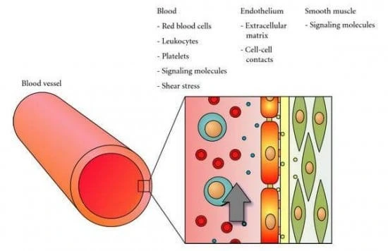
Endothelial cell image from Freya Mowat (wellcome collection – CC BY 4.0), Channel drawing figure by Raasch et al. (2015), Vascular cells drawing figure by van der Meer et al. (2009).
Microfluidics endothelial cell culture
Endothelial cell layer culture in a microfluidic channel is now accessible using this pack for an in vitro model as close as possible to in vivo conditions. Our plug-and-play instruments are the easiest solution to transition from classical 2D cell culture in wells to organ-on-chip technologies.
Based on the high-accuracy OB1 flow controller (Elveflow) and a membrane biochip, this solution contains all the required microfluidic parts for researchers to set up their endothelial cell culture with improved EC marker protein expression and good cell adhesion. Our experts will also accompany you during the setup of the microfluidic platform.
Experiments involving organs-on-chips are complex systems to set up. Fortunately, we were engaged in several organ-on-chip projects over the years and gathered valuable knowledge in this research field. We are always eager to discuss ideas with researchers!
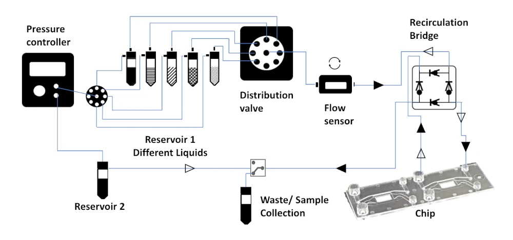
A basic microfluidic endothelial cell layer culture pack contains a pumping channel coupled with a distributor to seed two different kinds of cells on both sides of the membrane in the chip. This allows the creation of a more physiologically relevant endothelial cell layer that can be used for new therapeutic developments or toxicity screening.
The distributor quickly injects different substances, like FITC-dextran, and a 3/2 valve is used to choose which channel to perfuse. The efficiency of the perfusion will be directly linked to the flow rate inside the upper and lower channels. Flow rates can be measured with a microfluidic flow sensor (e.g., Galileo).
This setup allows for the control of strain, shear stress, and pressure to approach physiologically realistic values. Thus, this pack’s experiment conditions are more relevant than those of classical static cell culture.
Microfluidic endothelial cell culture setup
An all-in-one pack guarantees perfect compatibility between the different parts, allows a quick experiment start, is piloted by a single software, and can be used for other different applications. This is why a pack is the easiest way to set up your microfluidic experiment.
Setup
Flow sensor (Galileo, MIC)
Software (Galileo user interface)
Recirculation Bridge
Flow controller
Rotative valve
Cross-flow membrane chip from microfluidic ChipShop (Suggestion: Fluidic 568)
Pressure splitter manifold
Several falcon reservoirs for the sample and the medium
3/2 Microfluidic valves
Valve controller
Tubings and connectors
Primary human umbilical vein endothelial cells (HUVECs) (if necessary)
We have recently published a review comparing different perfusion recirculation systems.
Why use microfluidics for endothelial cell culture?
Firstly, using microfluidics is a way to decrease the amount of potentially valuable samples needed for a reaction. Moreover, fluid properties can be precisely tuned to mimic in vivo conditions at the microfluidic scale as much as possible.
Thus, creating microfluidics models of human organs is more effective than 2D classical or animal models because microfluidic systems allow more physiologically accurate conditions. Furthermore, the European Union and the public are pushing to reduce the use of animal models.
Microfluidic endothelial cell layers allow more flexible, precise, and efficient experiments to assess drug toxicity or pathogen effects on endothelial cells.
References
1. A. D. Van der Meer, A. A. Poot, M. H. G. Duits, J. Feijen, I. Vermes, “Microfluidic Technology in Vascular Research”, BioMed Research International, vol. 2009, Article ID 823148, 10 pages, 2009.
Microfluidics endothelial cell culture principle
Much work has investigated how endothelial cells react to various chemical, biological, or physical stimuli in vitro, as vascular dysfunction is a critical consequence of significant diseases like diabetes or cancer [1]. Organs-on-chip is an up-and-coming field that will prevent drug clinical failures and replace classical 2D cell culture and animal model testing [2].
Endothelial cell culture models can be created in microfluidic channels separated by a membrane to obtain physiologically relevant biomechanical conditions with realistic flow, strain, shear stress, and pressure [3-4].
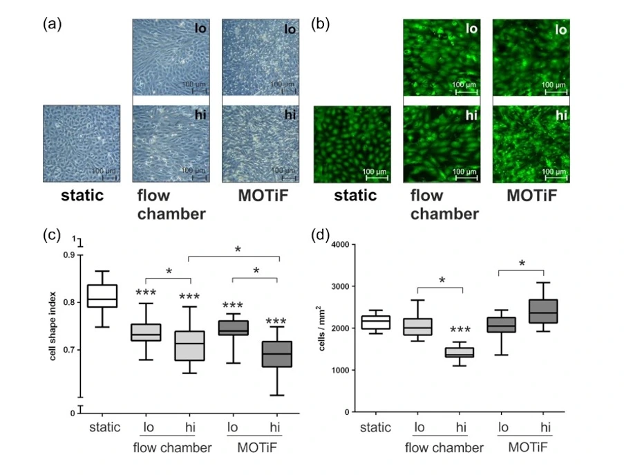
For example, Raasch et al. compared the viability and morphology of endothelial cell cultures in traditional static and dynamic conditions, with flow cells, and in a microfluidic system, as illustrated below.
The low flow conditions were 0.7 dyn cm−2 with only minimal flow-related mechanical stimulation and high shear stress with ten dyn cm−2, which corresponds to the human in vivo situation in larger arteries and venules and represents the mean shear stress rate of the common human carotid artery.
The authors have not found a significant difference in cell viability between low-flow conditions. However, cells presented better elongation and density in the high-flow condition when inside the microfluidic chip, indicating better adaptability [5].
This kind of microfluidic system has also been used to study the endothelial cells at the blood-brain barrier to make a human-relevant disease model [6]. The permeability of the endothelial cell layer in function of the shear stress applied on the layer has also been studied in a two channels system separated by a membrane [7].
Check out our other organ-on-a-chip packs: blood-brain barrier on-a-chip, gut-on-a-chip, bone on-a-chip and lung-on-a-chip.
References
A. D. van der Meer, A. A. Poot, M. H. G. Duits, J. Feijen, I. Vermes, “Microfluidic Technology in Vascular Research”, BioMed Research International, vol. 2009, Article ID 823148, 10 pages, 2009
Capulli A. K., Tian K., Mehandru N., Bukhta A., Choudhury S.F., Suchyta M., Parker K.K., Approaching the in vitro clinical trial: engineering organs on chips, Lab Chip, 2014,14, 3181-3186
Estrada R., Giridharan G.A., Nguyen M-D, Roussel T-J, Shakeri M, Parichehreh V., Prabhu S.D., and Sethu P., Endothelial Cell Culture Model for Replication of Physiological Profiles of Pressure, Flow, Stretch, and Shear Stress in Vitro, Anal. Chem. 2011, 83, 8, 3170–3177
Estrada R., Giridharan G.A., Nguyen M-D, Roussel T-J, Prabhu S.D., and Sethu P., Microfluidic endothelial cell culture model to replicate disturbed flow conditions seen in atherosclerosis susceptible regions, Biomicrofluidics, 5, 032006 (2011)
Martin Raasch et al. Microfluidically supported biochip design for culture of endothelial cell layers with improved perfusion conditions, 2015, Biofabrication 7 015013
L. M. Griep, F. Wolbers, B. de Wagenaar, P. M. ter Braak, B. B. Weksler, I. A. Romero P. O. Couraud, I. Vermes & A. D. van der Meer, A. van den Berg, BBB ON CHIP: microfluidic platform to mechanically and biochemically modulate blood-brain barrier function, Biomedical Microdevices volume 15, 145–150 (2013)
Young E. W. K., Watson M. W. L., Srigunapalan S., Wheeler A. R., Simmons C. A., Technique for Real-Time Measurements of Endothelial Permeability in a Microfluidic Membrane Chip Using Laser-Induced Fluorescence Detection, Anal. Chem. 2010, 82, 808–816
Customize your pack
The cross-flow membrane chip included in this pack can be in Cyclo Olefin Copolymer (COP) or Polystyrene (PS), hydrophilized or not. Depending on your application, two different pore sizes can be chosen: 0.2 µm or 8 µm. Custom-specific membranes can also be made on demand.
This pack can be significantly personalized depending on your requirements.
Bubbles can be a problem for cell culture. Thus, we can provide a bubble remover to tackle this issue.
Other packs for organ-on-chips are available. Check out the blood-brain barrier on-a-chip, gut-on-a-chip, bone on-a-chip, and lung-on-a-chip.
You can contact our scientists to answer any questions about this endothelial cell layer culture pack and how it can match your applications.
Frequently Asked Questions
Can I order a pack?
A pack can be available under certain conditions. Since packs are products that are still being developed, we have a few eligibility criteria to maximize their success rate. We need to discuss your specific needs with our experts to provide a personalized response.
Can the pack be put inside the CO2 incubator?
Some of our instruments can be placed inside the CO2 incubator. Depending on your needs, our team will be able to suggest the best configuration and include incubator-friendly equipment.
Can a pack be customized based on my specific application?
Yes! Our experts will establish which instruments are best suited for your application, such as the type of flow sensor or the number of flow controller channels you need to perform your experiment. Contact us using the “talk to our experts” green button above.
Can I buy individual instruments?
Our instruments are in the beta testing phase and can be tested as a pack or individually, so get in contact with our team to know how our beta testing program works.
Download the MIC Horizon Europe 2026/2027 Calls Calendar:
Products & Associated Accessories
FAQ - Endothelial Cell Culture Pack
What is the actual concept of Endothelial Cell Culture Pack?
An endothelial cell culture pack is a biologically active cellular monolayer with stable hemodynamic forces. It is used to automate endothelial cell culture and improve result reproducibility with a more physiologically relevant microenvironment.
Is it perfectly suitable for the long-term experiments?
Yes. The system can sustain hemodynamic stresses for a longer trial duration, making it appropriate for protracted cultivation. Because of this, it is beneficial for research where continuous biomechanical stimulation and consistent perfusion are crucial.
Can the pack be customized for specific applications?
Yes. The pack can be greatly customized. Users can request unique membranes, add features like a bubble remover, specify pore diameters of 0.2 µm or 8 µm, and select membrane chip materials like COP or PS. Additionally, according to the business, it can assist in choosing the appropriate type of flow sensor, the number of controller channels, and configurations that are suitable for incubators.
How microfluidic endothelial cell culture system operate?
In a straightforward implementation of the pack, two different cell types are sown on two sides of a membrane within the chip with the help of a pumping channel and distributor. The distributor may select the channel to perfuse by a 3/2 valve, and the quick injection of materials such as FITC-dextran can be done. The flow rate in the upper and lower channels is one of the critical factors that can be measured with a microfluidic flow sensor.
What are the main benefits of this pack for researchers?
A single software solution operates the pack, ensuring component compatibility and enabling rapid startup. Additionally, the arrangement can increase the expression of endothelial cell marker proteins and cell adhesion and offers clients more physiologically appropriate culture-conducive conditions than stagnant culture techniques.
Why use microfluidics for endothelial cell culture instead of conventional static culture?
Microfluidics allows for better control of the fluid’s properties to mimic real body conditions while using fewer samples. Endothelium micromodels are more helpful in medication toxicity studies, vascular research, and pathogen-response trials because they can replicate realistic flow, pressure, strain, and shear stress more accurately than traditional 2D culture.
What components are included in the pack?
The following is the list of the components typically included in the pack:
- OB1 Flow Controller
- Membrane Biochip
- Flow Sensor
- Control Software
- Rotary Valve
- Cross-Flow Membrane Chip
- Pressure Splitter Manifold
- Reservoirs
- 3/2 Microfluidic Valves
- Valve Controller
- Tubing
- Connectors
- Primary Human Umbilical Vein Endothelial Cells (HUVECs) (optional)
What kinds of applications is this pack useful for?
The pack aims to create endothelium models that are more physiologically appropriate, thereby facilitating toxicity screening and treatment development. Additionally, investigations of endothelial reactivity to flow conditions, permeability, and disease-relevant microenvironments, including the blood-brain barrier, are highlighted on the page along with more general organ-on-a-chip and vascular research use cases.
Still have some questions? Get in touch with our experts to learn how this solution can support your research.
