Bone-on-a-Chip Pack
Easily mimic the complex bone physiology and extracellular environment
Control complex microenvironments
More relevance and controllability than traditional models
Up to 3-week long cell cultures
Continuous and controlled supply of nutrients in a stable flow
Fail-safe mechanism
Stop losing your cell experiment due to clogging
Plug-and-play platform
Beginner friendly pack with detailed user guide

Need a microfluidic SME partner for your Horizon Europe project?
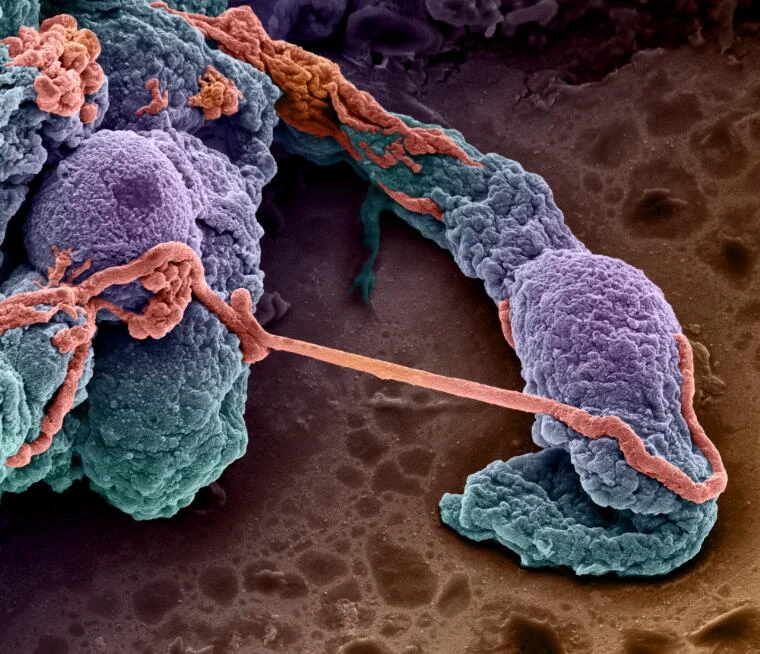
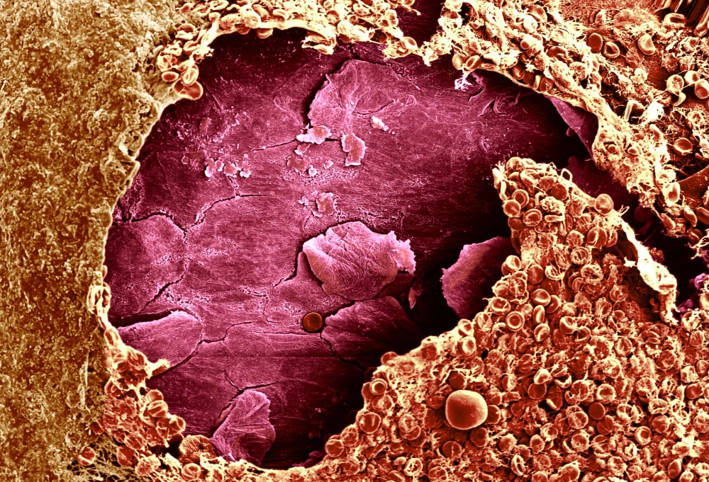
Figure 1. SEM bone, rotten trabeculae (osteoporosis). Credit: David Gregory & Debbie Marshall. Attribution 4.0 International (CC BY 4.0).
Bone-on-a-chip advantages over traditional models
Mimic in vivo complex microenvironment
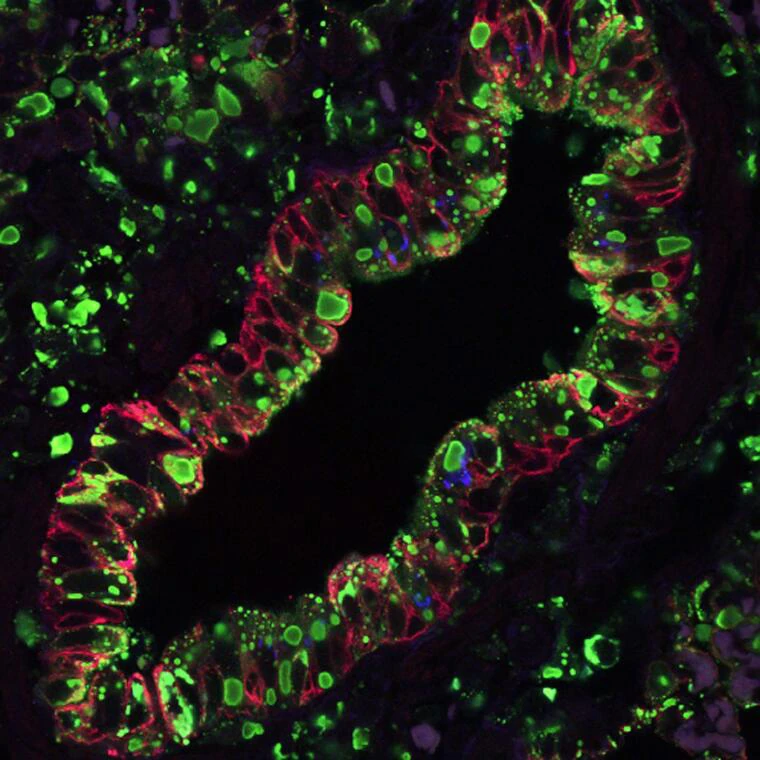
The bone is one of the most complex organs in the body. It operates in a fine balance between osteocytes and osteoclasts, with the former synthesizing new bone and the latter resorbing it [1]. Therefore, cell-to-cell interaction, either through ligand-receptor binding or ionic gradients, is a major component of bone physiology.
However, reproducing it in traditional cell culture models is challenging because of the large volumes and dimensions of the systems, besides being poorly controlled. Culturing bone cells on a chip address precisely these limitations.

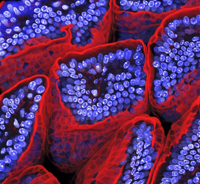
Bone. Credit: Kevin Mackenzie, University of Aberdeen. Attribution 4.0 International (CC BY 4.0).
Modulate the extracellular matrix
Bone cells are organized around osteons, cylindrical structures parallel to the longitudinal axis of the bone, housing blood vessels and innervations [1].
They are hard to reproduce in traditional cell cultures but are easily integrated into microfluidic chips, such as the Pillar Chips (Fluidic 261) of ChipShop, increasing the morphological relevance of the model.
References
1. Mansoorifar, A., Gordon, R., Bergan, R. C., Bertassoni, L. E., Bone-on-a-Chip: Microfluidic Technologies and Microphysiologic Models of Bone Tissue. Adv. Funct. Mater. 2021, 31, 2006796. https://doi.org/10.1002/adfm.202006796
Bone-on-a-chip setup
We have assembled all the components to perform a successful bone cell culture on a chip. The setup displayed below has two main functions: 1) the continuous and stable perfusion and recirculation of media for a highly controlled microenvironment; 2) the fail-safe mechanism to ensure the preservation of the experiment in case of clogging. Also, the bone-on-a-chip Pack can be adjusted according to your application.
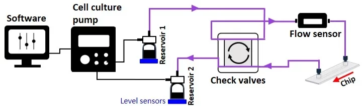
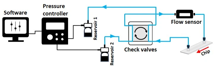
The bone-on-a-chip pack contains:
Flow sensor (Galileo, MIC)
Level sensors
Software (Galileo user interface)
Flow controller
Valves
Fittings, tubings & luers
Reservoirs
Microfluidic chip for bone cell culture (suggestion: ChipShop’s Fluidic 261)
User guides for instruments
This pack can also be adapted to be used with our automated cell culture platform.
In case of applications requiring fluid injection to test the effects of drugs, for example, a rotary valve can be added to the setup.
Bone-on-a-chip applications
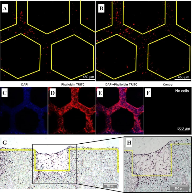
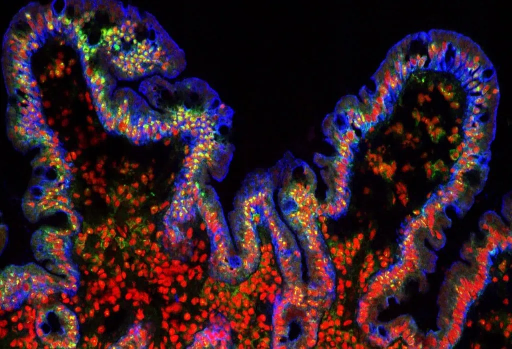
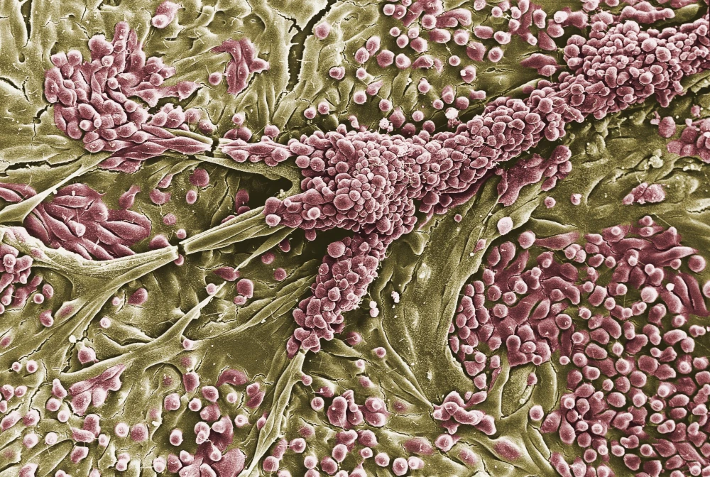
To investigate osteogenesis, Bahmaee et al. designed a microfluidic bone-on-a-chip with pillars made of a functional material, PolyHIPES [1]. The authors cultured human embryonic stem cell-derived mesenchymal progenitor cells (hES-MPs) for 3 weeks and tested different flow rates and profiles to assess metabolic activity, osteogenic differentiation, and mineralized matrix deposition.
Their results show the influence of shear stress on gene expression and cellular behavior. For example, an intermittent flow profile promotes cell differentiation and enhances mineralized matrix deposition. The functionalized material of the microfluidic chip was also shown to be appropriate, with cells interacting with the surface and covering its full extent, as shown below.
Our placenta model platform can be used as a barrier model to study molecular transport in other applications, such as:
Gut-on-a-chip pack
Intestinal cells coculture under flow, mimicking the gut physiology
✓ All microfluidic pieces included, quick and easy assembly
✓ Dynamic culture conditions
✓ Advanced in viro/ex vivo
Gut-on-chip
Inflammatory bowel disease model
Automatically collect important markers of IBD in a relevant in vitro model
✓ Uncover cytokine profile changes in time
✓ Mimic pathological conditions of IBD
✓ Tailor sample volume to your analysis
Blood-brain Barrier on Chip
Plug-and-play instrument pack for long term BBB on a chip study
✓ Relevant microenvironment
✓ Automatized organ-on-chip perfusion
✓ Plug-and-play microfluidic platform
Blood-brain Barrier on Chip
Liver-on-a-chip model
Mimic the liver microenvironment in long term experiments
✓ Improve your reproducibility with physiological culturing conditions
✓ Automated and controlled supply of nutrients in a stable flow
✓ Test different conditions at the same time
Liver-on-chip
Lung-on-a-chip model pack
Perform lung research in a physiologically relevant microenvironment
✓ Culture your lung cells in a physiological air-liquid interface
✓ Continuous and controlled supply of nutrients in a stable flow
✓ Stop losing your cell experiment due to clogging
Lung-on-a-chip
Skin-on-a-chip
Reproduce the dynamic extracellular environment of the skin with ease
✓ Compatible with air-liquid interfaces
✓ Compatible with teer measurements
✓ Compatible with live cell imaging
Skin-on-a-chip
Kidney-on-a-chip
Physiological microenvironment for a more realistic in vitro kidney model
✓ Physiological flow rates
✓ Use the chip of your preference
✓ Enrich the media with metabolites
Kidney-on-a-chip
We have recently published a review of the different organ-on-a-chip models and current innovations.
References
1. Bahmaee H, Owen R, Boyle L, Perrault CM, Garcia-Granada AA, Reilly GC and Claeyssens F (2020) Design and Evaluation of an Osteogenesis-on-a-Chip Microfluidic Device Incorporating 3D Cell Culture. Front. Bioeng. Biotechnol. 8:557111. doi: 10.3389/fbioe.2020.557111
Customize your pack
Our packs are highly customizable, so you can use the extra flexibility to adapt them to your specific needs. Our microfluidic specialists will advise you on the best instruments and accessories depending on your needs and will accompany you during the setup of the microfluidic platform.
Frequently asked questions
Can I order a pack?
Since Packs are products that are still being developed, we have a few eligibility criteria to maximize their success rate. A discussion with our experts is needed to determine your specific needs to offer you a personalized response.
Is the setup sterilizable?
Yes, we have developed a simple protocol for sterilization and cleaning that is provided along with the user guide.
Can a pack be customized based on my specific application?
Yes! Our experts will establish which instruments are best suited for your application, such as the type of flow sensor or the number of flow controller channels you need to perform your experiment. Contact us using the “talk to our experts” green button above.
Can I buy individual instruments?
Our instruments are in beta testing phase and can be tested as a pack or individually, so get in contact with our team to know how our beta testing program works.

Funding and Support
The LIFESAVER project helped develop this pack. This project is funded by the European Union’s H2020-LC-GD-2020-3, grant agreement No. 101036702 (LIFESAVER).
Products & Associated Accessories
FAQ - Bone on a Chip Pack
What is a bone-on-a-chip, and why would it be required?
The bone-on-a-chip is a microfluidic system that mimics the in vitro physiological microenvironment of bone tissue in a continuous, regulated perfusion. This is required because the bone is one of the most complex organs in the body, functioning through a fine balance between osteocytes, which form new bone, and osteoclasts, which break it down. Such a dynamic and interactive environment is extremely difficult to reproduce in traditional cell culture systems, which are large in volume, poorly controlled, and incapable of maintaining the cell-to-cell interactions central to bone physiology.
What are the biological structures that this model is intended to model?
The organisation of bone cells is around osteons, cylindrical structures parallel to the bone’s longitudinal axis that harbour blood vessels and nerve fibres. The structures regulate local nutrient delivery, mechanical signalling, and ionic gradients across cell types. Easy to grow in standard culture formats, they are naturally incorporated into pillar-based microfluidic chips, such as the Fluidic 261 from ChipShop, which more closely resembles the architectural geometry of these units of the osteon, thereby significantly enhancing the morphological relevance of the model.
What are the two fundamental functions of the bone-on-a-chip setup?
The platform is developed based on two main functions. To begin with, it ensures stable, continuous perfusion and recirculation of culture media, thereby maintaining a tightly controlled microenvironment during the experiment. Second, it also has a fail-safe mechanism, which helps prevent the experiment from clogging, which is a frequent failure mode in long-duration microfluidic cell cultures, so that weeks of effort are not wasted in case of a single blockage in the fluidic circuit.
What is contained in the bone-on-a-chip pack?
The pack itself includes a flow sensor (Galileo, MIC), level sensors, a flow controller, valves, fittings, tubing and luer connectors, reservoirs, the Galileo user interface software, user instructions on each of the instruments, and a microfluidic chip, compatible with bone cell culture – the Fluidic 261 Pillar Chip of ChipShop will be the default choice. Everything is compatibility tested, and can be configured in a plug-and-play configuration, available even to microfluidics scientists unfamiliar with it.
What is the role of shear stress in bone cell culture on chip?
The mechanical force applied to cells by moving media, known as shear stress, has been reported to affect gene expression and cellular behaviour in bone cultures. In a study involving human embryonic stem cell-derived mesenchymal progenitor cells (hES-MPs) in three-week cultures, it was shown that the flow profile strongly influences osteogenic differentiation and mineralized matrix deposition. It is important to note that an intermittent flow profile was demonstrated to favour cell differentiation and greater matrix mineralisation compared to constant flow, highlighting the value of accurate flow direction in this model.
What is the maximum duration that cell cultures can be sustained on this platform?
The platform allows cell cultures of up to three weeks. The length of this period is made possible by the automated media recirculation system which supplies the nutrients to the chip continuously and ensures the conditions of stable flow. The built-in fail-safe clogging prevention system also ensures that experiment integrity is maintained throughout this duration thus minimising the possibility of surprise culture loss as is common in long-duration microfluidic systems.
Is it possible to use the pack to do drug testing or inject compound?
Yes. A rotary valve may be introduced into the setup to allow the use of drugs or test compounds when there is a need to introduce them to an ongoing culture. This option enables the researcher to inject certain substances at set time points throughout the experiment without interfering with the continuous perfusion, which makes the platform appropriate in pharmacological studies, tests of toxicity and studies of the role of drugs on bone cells behaviour or matrix composition over time.
Does it support other MIC platforms or instruments?
Yes. The bone-on-a-chip pack can be extended to the automated cell culture platform offered by MIC that offers even greater degree of integration and automation. It is also made in the same modular Galileo instrument ecosystem as the other organ-on-chip packs of MIC; this implies that those researchers that are already accustomed to the flow sensor and software backbone can easily switch bone culture experiments without requiring an extensive learning curve.
Is it possible to sterilise the setup between experiments?
Yes. This pack has a specially designed sterilisation and cleaning procedure, which is also included with the user manual. This is necessary to allow the system to be decontaminated safely between experimental runs, a requirement to preserve cell culture integrity across successive applications and/or in other applications where the control of contamination must be paramount, e.g. in long-term studies of osteogenesis or in experiments with primary patient-derived cells.
How do I get the pack, or tailor it, to use in my application?
The pack can be made extremely customisable, and our microfluidic experts will recommend the most suitable instrument configuration depending on the objectives of your research, the type of flow sensor used, the number of channels of the controller, the choice of chip, and any other modules, like a rotary valve to inject drugs. Since the pack is in beta testing, a preliminary discussion with our team is required before placing an order. It is also possible to access individual instruments using the beta programme.
