Patented technology
Grooved Microscope Slides for Liquid Samples
Micropatterned microscope slides for liquid handling
Highly controlled environment
Highly controlled extracellular environment with stable flow
Compatible with CO2 incubator
Adapted to standard CO2 incubators and biosafety hoods
Up to 4 independent cell cultures
Save time and improve reproducibility
Up to 3-week cell cultures
Less manual work and more accuracy

Need a microfluidic SME partner for your Horizon Europe project?
Microscopy with liquid samples
In many areas of research, detecting particles, called “particles of interest,” in a liquid is crucial. Researchers might also study how these particles behave over time or even try to encourage them to form new structures. These particles can include biological entities like cells (animal or plant, living or not), antibodies, proteins, or viruses. They can also be non-biological, like certain chemical molecules, polymers, or fluids. It’s all about understanding how these particles interact and evolve within the liquid.
Most sample analysis protocols involve using a glass slide and an observation tool, like a microscope or spectrum analyzer. The sample is placed on the slide, and a coverslip is gently added on top to avoid trapping air bubbles or damaging any particles. The slides stay in place due to capillary action, and in some cases, the sample is glued to the slide (often with wax) to prevent evaporation, especially during long observations or when the temperature changes.
Limitations of using regular microscope slides
This type ofprotocol comes with several challenges. When liquid is placed on the slide, it often spreads uncontrollably into a thin film just a few microns thick. This can waste samples along the edges and make it hard to control where theliquid spreads, especially if the analysis instrument requires a specific zone.
Adding the top coverslip can also be challenging. Capillary forces may pull the liquid towards one edge of the slide, wasting more sample and making it harder to keep it centered. The liquid can also spread unevenly, leading to inconsistent thickness that’s difficult to manage.
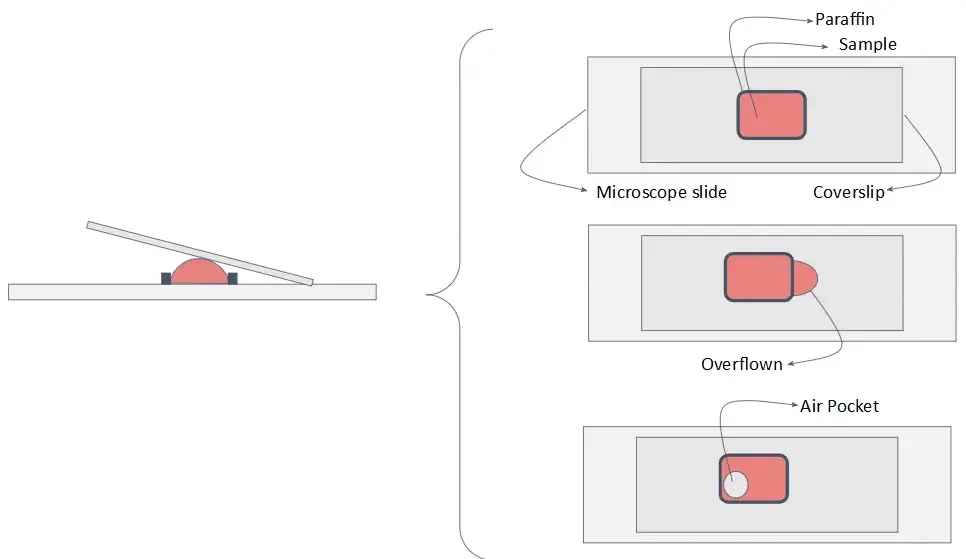
Finally, any liquid at the slide’s edges can interfere with sealing the system during sealing, making the process dependent on the user’s skill. This variability often results in inconsistent sample preparation and unreliable analysis results. Liquid outside the slide can also cause issues when trying to create a precise, thin cavity for the sample.
The presence of liquid outside the slide and/or coverslip, which may come into contact with the user and/or the analysis instrument, is generally considered unacceptable, particularly in the case of samples containing carcinogenic or toxic products. Moreover, the sealing step is particularly time-consuming to implement and requires the intervention of an experienced user.
Microscope slides for liquid samples
With these limitations in mind, we specifically designed a microscope slide to handle liquid samples. It consists of a glass slide with engraved micropatterns or grooves around the sample visualization area. These micropatterns, which act as capillary barriers, are continuous and help contain the liquid within the area of interest without the formation of air bubbles.
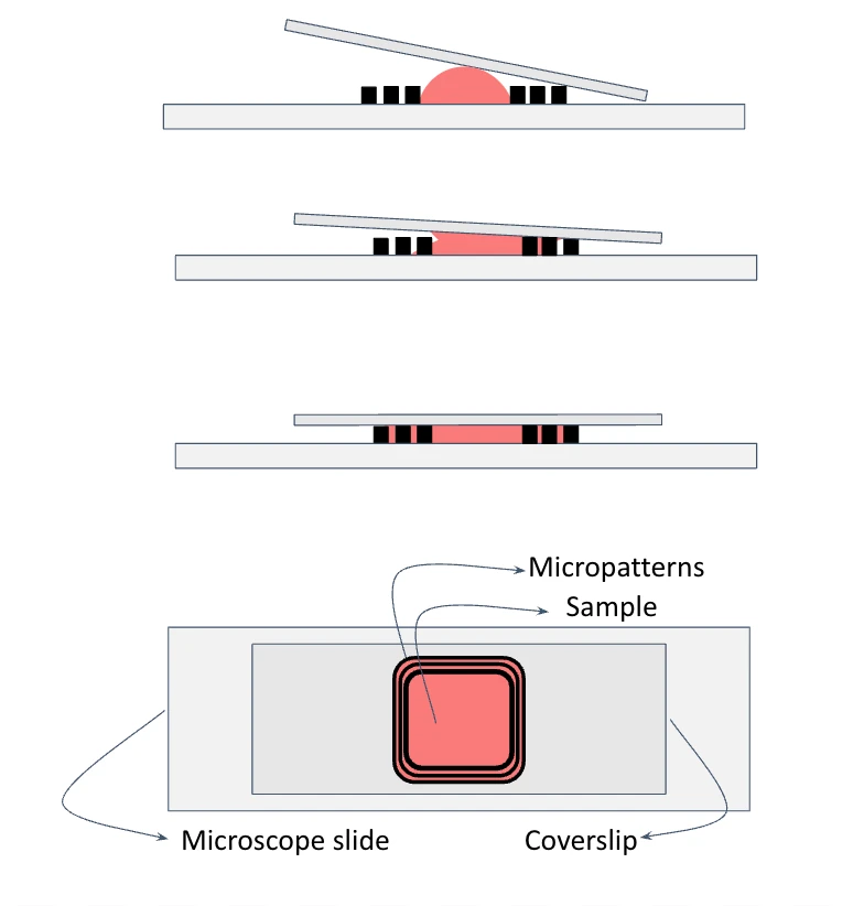
The surface of the glass slide and the grooves can be modified to better host different sample types.
References
Featured microscope image: Author: Morgan Brown, CSIRO – http://www.scienceimage.csiro.au/image/11403
Patent
(EN) LIQUID SAMPLE SUPPORT SUBSTRATE, ASSEMBLY COMPRISING SUCH A SUBSTRATE AND USE THEREOF
(FR) SUBSTRAT DE SUPPORT D’ÉCHANTILLON LIQUIDE, ENSEMBLE COMPORTANT UN TEL SUBSTRAT ET SON UTILISATION
Other applications
Some biological applications of our microscope slides also include:
Microscopic observation of biological samples
Detection of molecules of interest
Chemical reaction chamber
Spectrometry analysis
And many more!
Microscope slides for liquid samples
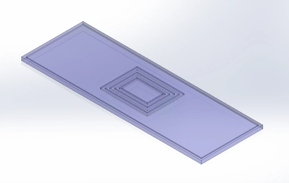
The technical specifications of the microscope slides for liquid samples are:
| Technical Specifications | |
|---|---|
| Wetted materials | Glass or thermoplastics (PVD, CVD, PECVD) |
| Dimensions | Micropatterns:
Distance between micropatterns: 100 µm |
| Working volumes | TBD |
Customize your pack
Our instruments can be added to different setups depending on your specific needs. In this light, our microfluidic specialists will advise you on the best instruments and accessories depending on your needs and will accompany you during the system’s setup.
Frequently asked questions
Can the working volume or micropatterns of the microscope slides be altered?
For sure! For more information on custom designs, contact us at innovation[at]microfluidic.fr or just click on the “talk to our experts” green button on top.
Are the microscope slides also compatible with upright microscopes?
Yes, by using a conventional coverslip, you can image your sample with inverted or upright microscopes without problems.
Are the microscope slides reusable or disposable?
We recommend to dispose and use new microscope slides for each experiment, but you can clean the slides and reuse them if you wish.
Products & Associated Accessories
FAQ - Grooved Microscope Slides for Liquid Samples
What are grooved microscope slides?
Grooved microscope slides are glass slides with carved micropatterns specifically designed to handle liquid samples. It consists of a glass slide with engraved micropatterns or grooves around the sample visualization area. These micropatterns, which act as capillary barriers, are continuous and help contain the liquid within the area of interest without the formation of air bubbles.
What problems with conventional slides do these address?
The following are some of the common problems associated with standard microscope slide protocols:
- Liquid waste sample material spread unpredictably, creating a thin, uncontrollable film.
- The pressure between capillaries can cause the liquid to be offset when a coverslip is used, making homogeneous analysis difficult; the uneven sample thickness further complicates this.
- The lack of liquid flowing out at the slide is a safety concern in toxic or carcinogenic samples, and sealing the slide edges is tedious and requires skill.
The grooved design solves these issues by functioning as a passive containment system.
What types of samples are these slides suited for?
The slides are used for the following types of samples & applications :
- Biological samples: including antibodies, proteins, viruses, live or fixed cells (plant or animal).
- Chemical samples: fluid systems, polymers, and reactive molecules.
- Applications: Microscopic observation of biological samples, Detection of molecules of interest, Chemical reaction chamber, & Spectrometry analysis
Are the slides compatible with standard microscopy setups?
Yes. The slides can be used with both a stand and an inverted microscope. The samples are slide-covered with a standard coverslip for observation under the upright microscope. The slides can also be used in biosafety hoods and regular CO2 incubators; therefore, this makes the slides suitable in live-cell imaging operations.
Can the micropattern dimensions or working volumes be customized?
In fact, they can be custom-designed; i.e., modified working volumes, groove sizes, and pattern geometries. Request a quote on the product page to provide specifications.
Can the slide be reused?
It is recommended to use single-use per experiment to ensure repeatability and avoid cross-contamination. However, assuming that your process allows it, the slides may be reused and cleaned.
What are the main advantages of applications using cell culture?
The slides can maintain cultures for up to 3 weeks and can carry up to 4 distinct, parallel cell cultures. Among the key benefits, there are:
- Regulated extracellular environment under constant flow
- Reduced the number of manual interventions and handling
- Better reproducibility between experiments
- It is compatible with long observations with no evaporation problems.
Is this system protected by intellectual property?
Yes. A micropattern design was registered as an international patent, WO2017042115, for a liquid sample support substrate, an assembly comprising such a substrate, and its use.
