Neuron Culture Pack - Low Shear Cell Culture
Cell culture system for shear-sensitive cell lines
Safe neuron culture under flow
Advantages of dynamic cell culture even with shear-sensitive cells
Highly controlled microenvironment
Continuous and controlled supply of nutrients in a stable flow
Up to 3-week cell cultures
Less manual work and more accuracy for experiments
Need a microfluidic SME partner for your Horizon Europe project?
Neuron culture pack
Neuron culture and shear stress
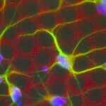
Unlike other cells in the body, the physiological microenvironment of neurons is usually in tissues that do not have continuous fluid flow. Therefore, neuron culture under media perfusion, exposed to shear stress, can significantly affect their viability [1].
However, cell perfusion has marked advantages that can benefit neuron cell culture. For example, gradients can be formed to study the orientation of axons [1], or chemical cues can be precisely added to induce neural stem cell differentiation [2, 3]. On the other hand, the continuous flow of media ensures a constant supply of nutrients and waste removal, improving the microenvironment conditions [4].
Flow rate and shear stress
Shear stress is a direct effect of flow rate and system dimensions. For a system with defined dimensions, the lower the flow rate, the lower the shear stress. However, there is a limit to how low one can go with the flow rate and still have a stable flow. Thus, to have a system with a steady flow and low shear stress, it is necessary to re-think the dimensions and geometry of the culturing chamber.

Low shear bioreactor for neuronal culture
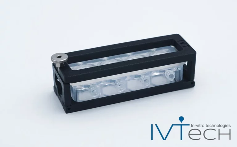
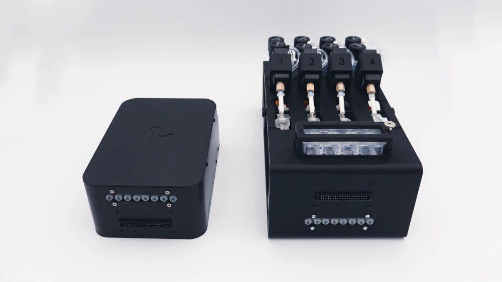
The Microfluidics Innovation Center partnered with IVTech, a biotechnology company specializing in advanced cell culture chambers, to refine in vitro models. MultiDyn has been designed and produced by IVTech SRL, an evolution of the LiveBox1 chamber. The system is a rack of 4 wells in parallel, compatible in dimension with the column of a standard 24-well plate.
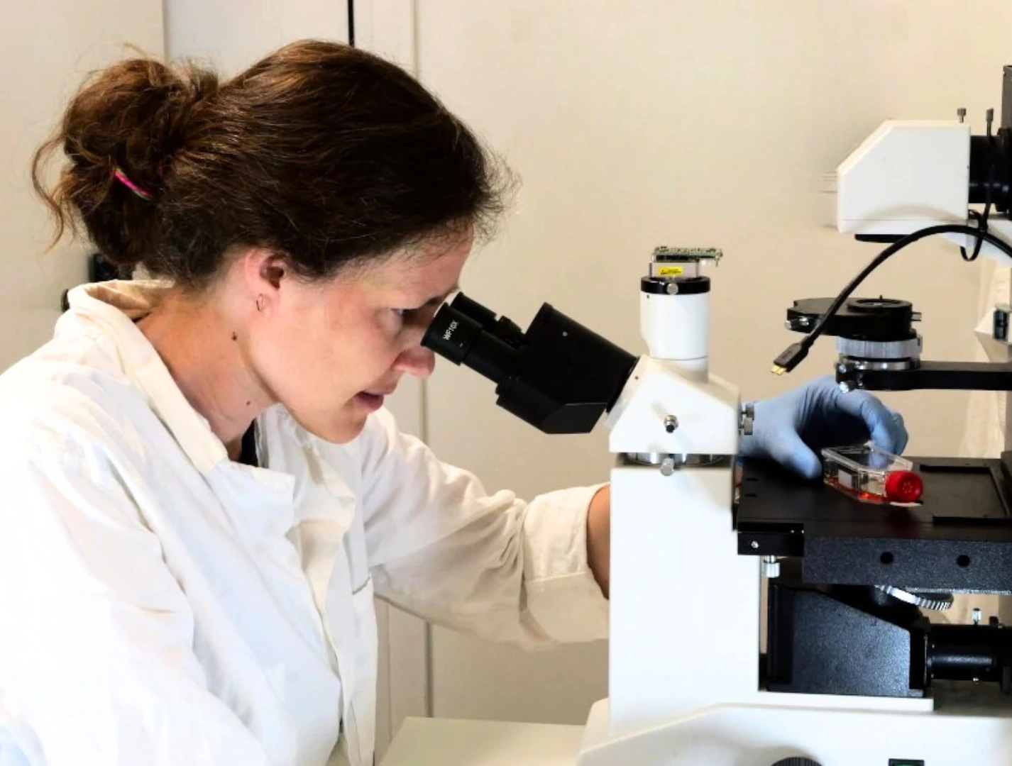
MultiDyn assures the opportunity to recreate all the experiments performed with LiveBox1, replicating the data in 4 parallel tests and reducing the complexity of the chamber setup. The rack is monitorable in real-time, using an inverted microscope, assuring the possibility of developing a 3D in-vitro model in dynamic conditions. Moreover, two or more racks can be joined in series to establish a multi-organ model, where each tissue communicates with the others due to a liquid exchange.
References
1. C. Joanne Wang, X. Li, B. Lin, S. Shim, G. L. Ming, and A. Levchenko, ‘A microfluidics-based turning assay reveals complex growth cone responses to integrated gradients of substrate-bound ECM molecules and diffusible guidance cues’, Lab Chip, vol. 8, no. 2, pp. 227–237, Jan. 2008.
2. T. Barra et al., ‘Neuroprotective Effects of gH625-lipoPACAP in an In Vitro Fluid Dynamic Model of Parkinson’s Disease’, Biomed. 2022, Vol. 10, Page 2644, vol. 10, no. 10, p. 2644, Oct. 2022.
3. B. G. Chung et al., ‘Human neural stem cell growth and differentiation in a gradient-generating microfluidic device’, Lab Chip, vol. 5, no. 4, pp. 401–406, Mar. 2005.
4. M. L. Coluccio et al., ‘Microfluidic platforms for cell cultures and investigations’, Microelectron. Eng., vol. 208, pp. 14–28, 2019.
Low shear and automated cell culture pack
The Microfluidics Innovation Center and IVTech combined their strengths to develop an automated pack for low-shear neuron cell culture. We used our flow control expertise to create an automated and secure cell perfusion system, integrating IVTech’s bioreactor for low-shear cell culture.

The neuron culture pack includes:
Automated cell culture platform comprising
Flow sensor eg. Galileo
Recirculation loops
Level sensors
Software (Galileo user interface)
Flow controller
IVTech MultiDyn bioreactor
User guide
Neuron culture in low-shear bioreactors
Barra et al. [1] used IVTech’s LiveBox 1 bioreactors to create an in vitro Parkinson’s disease (PD) model based on a neuron cell culture. They cultured a neuroblastoma cell line, SH-SY5Y, differentiated with retinoic acid as a model for P. Then, they tested and demonstrated the neuroprotective effects of a neuropeptide (pituitary adenylate cyclase-activating polypeptide, PACAP) in a fluid dynamic model.
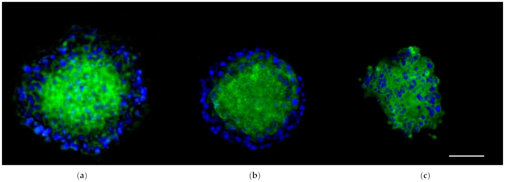
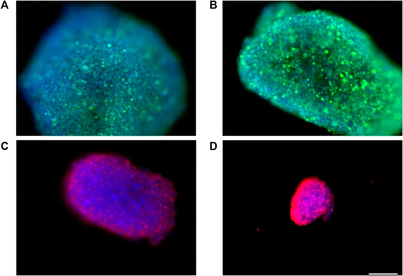
In another study to develop a blood-brain barrier model, the neuronal culture aimed to grow spheroids in the bioreactor [2]. According to the authors, “immunofluorescence experiments showed that fluid dynamic conditions have no appreciable influence on spheroids health, morphology, and organization.”
References
1. T. Barra et al., ‘Neuroprotective Effects of gH625-lipoPACAP in an In Vitro Fluid Dynamic Model of Parkinson’s Disease’, Biomed. 2022, Vol. 10, Page 2644, vol. 10, no. 10, p. 2644, Oct. 2022.
2. T. Barra et al., ‘gH625-liposomes deliver PACAP through a dynamic in vitro model of the blood–brain barrier’, Frontiers in Physiology, vol. 13. 2022.
Neuron culture pack specifications
The cell culture system comprises a perfusion platform and a pressure splitter. A flow controller controls the fluid flow.
The table summarizes the main specifications of the cell culture system.
| Components | Technical Specifications |
|---|---|
| Flow controller | 4 Channels (0 to 2000 mbar) |
| Microfluidic Flow sensor (Galileo) | Range from 0.5 to 10,000 µL/min |
| Recirculation loop | 4 check valves, polycarbonate, male Luer; compatible with 1/4″-28 standard microfluidic fittings |
| Pressure splitter | Valve controller with integrated Solenoid valves for compressed air. |
| Cell culturing chamber | MultiDyn bioreactor (4 parallel bioreactors, 1,5 ml internal volume, IVTECH SRL) |
The cell culture system is compatible with standard incubator conditions (37oC, 5% CO2, and 100% humidity); electronic components (except flow sensors) should be kept outside the incubator.
Customize your pack
Our Packs can be modified depending on your specific needs. Our microfluidic specialists will advise you to provide the best instruments and accessories, depending on your needs. They will accompany you during the setup of the microfluidic platform.
Frequently asked questions
Can I order a pack?
Since Packs are products that are still being developed, we have a few eligibility criteria to maximize their success rate. A discussion with our experts is needed to determine your specific needs to offer you a personalized response.
What can be placed inside the incubator?
The automated cell culture platform and the embedded flow sensors can be placed inside the CO2 incubator. The OB1 and the pressure splitter should be kept outside.
What type of cells can be cultured with the system?
Although shown here with a neuron culture, the system can be used to culture any cell type, as the culture media can be adjusted according to the cells’ needs, and the cell culturing chamber can be replaced by the required design.
Can the system be sterilized/autoclaved?
The components of the automated cell culture platform can be sterilized. Our user guide provides detailed information on how to do it.
Is the system compatible with microscopes/live cell imaging?
Yes, the MultiDyn bioreactors from IVTech are compatible with live cell imaging.


