Lymph Node Model Pack
Long term cell culture under flow outside the CO₂ incubator
Low flow rates
Get as low as 2 µl/min
Incubator independent setup
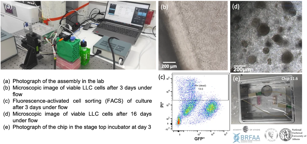
Culture cells up to 16 days outside the CO₂ incubator
Compatible with K-frame microscopes
The Stage top incubator was designed for live imaging

Need a microfluidic SME partner for your Horizon Europe project?
Lymph node model for lung metastasis
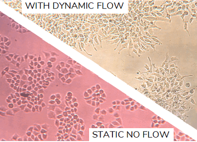
Developing complex models like this always brings researchers to the tough crossroads of choosing between the overly-complex and ethically-demanding use of animal models and the overly-simplistic and less-physiologically relevant in vitro models.
Now there’s an in-between. Culturing cell underflow improves the physiological relevance of fluid dynamic organs such as the lymph nodes while also allowing researchers to better isolate variables. In addition, the lymph node model is automatized for longterm cell culture outside the CO2 incubator, increasing its flexibility and portability and decreasing the manual labor and human error.
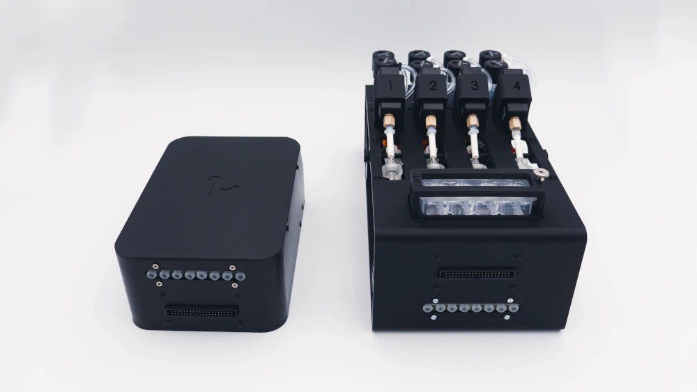
Setup
Cell culture pump
Flow sensor (Galileo, MIC)
Stage top incubator
Reservoirs
Tubings and fittings
Microfluidic chip
User guide
Software (Galileo user interface)
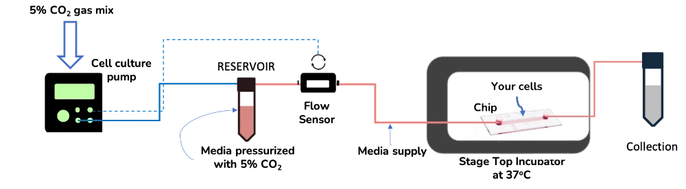
The setup includes the cell culture pump, which maintains the gas concentration fed into it to pressurize the liquid. For example, if a premixed bottle of 5% CO2 is plugged into the pump, mimicking a conventional CO2 incubator, it ensures that the media does not lose buffering CO2 to the atmosphere, so pH remains optimal for long periods outside the CO2 incubator. The flow sensor ensures that the flow is stable and reliable at a range of flow rates, starting at 2 µl/min.
The stage top incubator maintains the temperature at 37°C, but allows you to leave your cells on top of the microscope stage and perform live cell imaging with higher quality.
Reservoirs, tubing and connectors are all commercially available, can be reused or disposable, and can be bought independently and sterile. And the system was conceived to work with any type of chip, regardless if it’s commercial or home-made.
Lymph node model for lung metastasis in action
If you want to know more about organ-on-a-chip technology, have a look at this review!
References
- Gwóźdź, M. Pasieka-Lis, K. Kołodziej, J. Pankowski, R. Banaś, M. Wiłkojć, et al. Prognosis of patients with stages I and II non-small cell lung cancer with nodal micrometastases. Ann. Thorac. Surg., 105 (5) (2018), pp. 1551-1557.
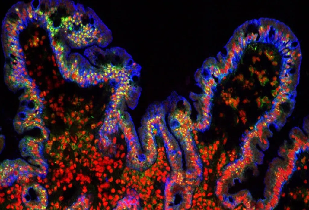
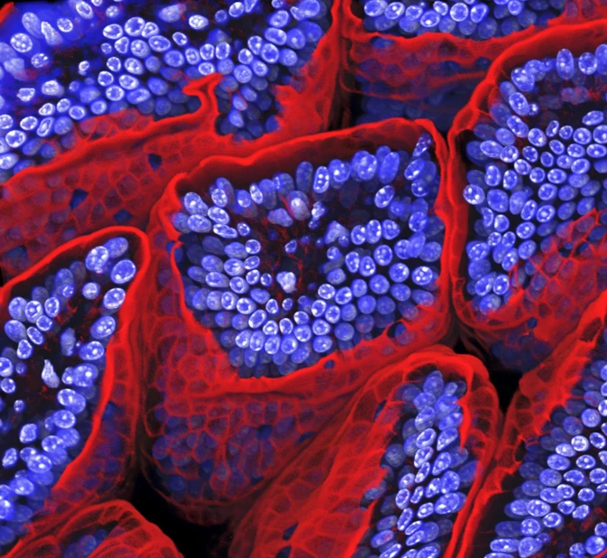
- Featured image description: This image of mouse lymph node was captured using multiphoton microscopy. B lymphocytes (red) accumulate inside B-cell follicle, while GFP-expressing myeloid cells (green) surround it. Collagen fibers (blue) run along lymph node conduits, capillary network (pink) delivers oxygen and nutrients to the follicle.
Caption: B-cell follicle inside a mouse lymph node
Wikimedia Commons, author Olena12
Compatibility and Applications
Some biological applications of our lymph node model pack include:
Cancer invasion and metastasis
Follow long-term cancer cell migration in real time on top of the microscope
✓ Keep ideal temperature conditions
✓ Follow cancer cell migration in real time
✓ Adapted to cultures under flow
Cancer cell migration
Cell-to-cell interactions
Metastasis on chip
And many more!
And the lymph node pack could also be adapted to other types of organs:
Gut-on-a-chip pack
Intestinal cells coculture under flow, mimicking the gut physiology
✓ All microfluidic pieces included, quick and easy assembly
✓ Dynamic culture conditions
✓ Advanced in viro/ex vivo
Gut-on-chip
Inflammatory bowel disease model
Automatically collect important markers of IBD in a relevant in vitro model
✓ Uncover cytokine profile changes in time
✓ Mimic pathological conditions of IBD
✓ Tailor sample volume to your analysis
Inflammatory bowel disease model
Blood-brain barrier on chip
Plug-and-play instrument pack for long term BBB on a chip study
✓ Relevant microenvironment
✓ Automatized organ-on-chip perfusion
✓ Plug-and-play microfluidic platform
Blood-brain Barrier on Chip
Liver-on-a-chip pack
Mimic the liver microenvironment in long term experiments
✓ Improve your reproducibility with physiological culturing conditions
✓ Automated and controlled supply of nutrients in a stable flow
✓ Test different conditions at the same time
Liver-on-chip
Lung-on-a-chip pack
Perform lung research in a physiologically relevant microenvironment
✓ Culture your lung cells in a physiological air-liquid interface
✓ Continuous and controlled supply of nutrients in a stable flow
✓ Stop losing your cell experiment due to clogging
Lung-on-a-chip
And many more!
Technical specifications for the lymph node model pack
The lymph node model pack is composed of two main pieces of equipment: the cell culture pump and the stage top incubator.
Cell culture pump technical specifications
The cell culture pump can come with 1 to 6 channels. Each channel connects to a flow sensor with a feedback-loop between pressure and flow rate. Each channel has the following specifications (tested with a set pressure value of 2 bar):
| Characteristics | Specifications |
|---|---|
| Accuracy | +/- 2.5 mbar |
| Air consumption | few mL/min |
| Response time | 140 ms |
| Settling time | 2750 ms |
| Overshoot | 0.12 mbar |
Stage top incubator technical specifications
The Stage Top Incubator was designed to allow cell culture on top of the microscope stage for live cell imaging.
| Characteristics | Specifications |
|---|---|
| Dimensions (mm) | 30.5 x 130 x 168 (h x w x l) |
| Base K- Frame | 3.5 x 110 x 160 (h x w x l) |
| Dimensions of internal usable space | 25 x 89 x 130 (h x w x l) |
| Dimensions of the bottom glass (ITO glass) | 1) 72 x 110 with a thickness of 1.1 mm 2) 50 x 25 with a thickness of 0.6mm 3) 50 x 22 with a thickness of 0.12 mm |
| Temperature range | Room Temperature to 70 °C |
| Temperature accuracy | ± 0,5 °C |
| External Material | Aluminum |
The stage top incubator is controlled by a converter connected to software (both included). Also, it is compatible with 1/16″ and 1/32″ ID tubing.
Frequently asked questions
Which gasses can the cell culture pump use for the lymph node model?
The cell culture pump can use any non-corrosive gas mixture.
Does the cell culture pump keep the gas sterile?
To guarantee the sterility of the used gasses, we advise adding a small disposable filter at the gas inlet of the reservoir to keep the sterile of your lymph node model.
Is the stage top incubator gas-tight?
No, the stage top Incubator allows gas exchange with the atmosphere.
What is the maximum flow rate that can be applied?
The system works well with the range of 0-5 ml/min.

Funding and Support
The Tumor-LN-oC project helped develop this instrument. This project is funded by European Union’s H2020-NMBP-TR-IND-2020, grant agreement-no. 953234 (Tumor-LN-oC).
Products & Associated Accessories
FAQ - Lymph Node Model Pack
What is this Lymph Node Model Pack?
It represents a commercially available microfluidic perfusion system that is designed to allow the long term culture of cells in flow and remains extracellular within a classical CO2 incubator. The basic premise is easy, maintain flow constant at very low-flow rates (even down to 2 ul/min), maintain pH steady by conditioning the gas with which the medium is pressurized, and maintain constant temperature on the microscope to image living cells.
What is the reason to use a lymph-node-like microfluidic model, rather than a typical in vitro model?
Lymph nodes are fluid-dynamic in nature: transport, gradients and cell traffic are important. Those effects are usually flattened by using static well plates whereas the use of animals can be slow, costly, and ethically cumbersome (not to mention more difficult to control variables). An example of this is a controlled microfluidic system, which is placed in between: you obtain dynamic perfusion, you have more control over conditions and you can measure the migration or interactions in real-time without ever having to guess what occurred between two endpoints.
What is it that is in the pack?
The pack is built around:
- A cell culture pump (pressure-driven, conditioning the gas stability (pH) with a gas)
- A microfluidic flow sensor (flow control with a feedback loop).
- Reservoirs
- Tubing and fittings
- A microfluidic chip (or compatibility guidance if you bring your own chip)
- A stage-top microscope-stage live imaging incubator.
- A user guide
- There is control software (Galileo user interface).
How does it keep the pH at incubator like conditions when the experiment is not in a CO2 incubator?
A cell culture pump pressurizes the culture medium with a given gas mixture. By connecting a premixed gas bottle (e.g., 5% CO2) the system will assist in preventing CO2 degassing the medium to ambient air, which is usually the source of pH drift in open and long experiments. Meaning: you are not actively modifying the chemistry in the buffer, it is just that you are avoiding the common loss of CO 2 that silently undermines long-term experiments.
What is the maximum time that I can sustain a culture?
The designed application is long-term culture, and it has been shown to work up to 16 days (under flow) in practice (the mechanics usually limits it to medium volume and nutrient depletion). If your endpoint is being limited by medium consumption, a recirculation strategy will allow you to continue experiments longer (and ensure that you do not fail in that this was working well until the reservoir was empty).
What is the range of the flow that the system is capable of supporting, and how stable is the system at low flow?
It has a low flow design and reaches down to 2 ul/min. At high flow rates, it performs well to approximately 5 ml/min. To achieve stability, it is used together with a flow sensor in a feedback loop (to keep the pressure constant to maintain the desired flow rate, rather than assuming the hydraulic resistance of the chip remains constant).
Which chips can I use? Should I have a particular proprietary equipment?
The design was developed such that it would be compatible with virtually any kind of microfluidic chip format- commercial or homemade as long as it has the capability to connect to standard tubing/fittings. It is frequently a huge issue in research consortia, as every lab has its chip geometry; a platform that one design might preclude any other geometry should the biology change.
Is it possible to perform live imaging on the microscope, and what formats of microscopes are supported?
Yes. The stage-top incubator was optimized to do live imaging on the microscope stage (can be configured to K-frame microscope formats as well). Cell culture is performed at 37°C yet you still retain you sample on the stage where you can view the sample in real-time to observe migration, invasion or cell-cell interaction.
What are the most common technical requirements of researchers (pump + incubator)?
Sanity-check specs are commonly provided in advance:
Cell culture pump (per channel; measured at constant pressure value of 2 bar):
- Accuracy: +- 2.5 mbar
- Response time: 140 ms
- Settling time: 2750 ms
- Overshoot: 0.12 mbar
- Air consumption: a few mL/min
Channel: The number of channels can be 1 up to 6 channels (each channel with a flow sensor to control it as a closed loop)
- Stage-top incubator:
- Operating temperature: Room temperature to 70 °C.
- Temperature accuracy: +- 0.5°C
- External material: aluminum
- Tubing compatibility 1/16 inch and 1/32 inch ID tubing.
Intended to be used on a microscope stage (with a pre-determined internal usable space, and with an option of a bottom glass, which is ITO glass).
What gases may the pump apply to, how do I maintain the gas path sterile?
Other non-corrosive gas mixture can be used in the pump. To achieve sterility, the simple suggestion is to include a small disposable filter to the gas inlet of the reservoir. It is the type of detail that is insignificant until you lose a 10-day running streak to contamination, and therefore is not a matter of option, but rather of requirement by most teams.
Which type(s) of research questions is this pack best suited to?
It glows when you are interested in dynamics: cell movement and invasion, cell-cell interactions, and behaviors associated with metastasis, and most other experiments where the time dependence is important, as well as the final point. It can as well be scaled to other organ-on-chip models (gut models, inflammatory disease models, blood-brain barrier, liver, lung-on-chip), provided that the chip and readouts can be compatible.
It is overkill when you just require a viability snapshot at 24-48 hours with no flow requirements- at this point, you are technically getting the same level of microfluidic control as you will need in the real experiment, but which is scientifically unnecessary.
Does the stage-top incubator have a gas-tight? Is it important to be concerned with gas exchange when having imaging?
No, it is not gas tight, it is permeable to the atmosphere. Practically, that is often tolerable (and even desirable) to make imaging convenient, but it does imply that you ought to consider your buffering plan and to think about how you are managing CO 2 and evaporation in regards to long periods of imaging time. The temperature stability on-stage is of interest to many groups and the pH stability up-stream is done through the conditioned pressurization gas and medium selection.
