Inline Viscosity of Cell Culture Media
Medium viscosity measured inline, at culture temperature.
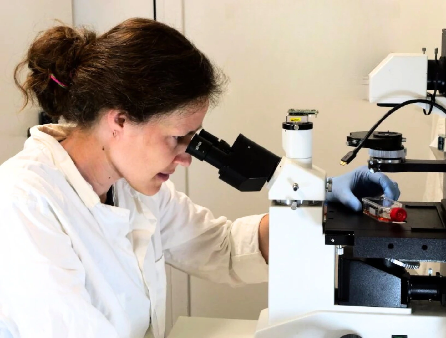
Reads shear-thinning media
Full viscosity curve (1-1000 s⁻¹) for accurate wall shear stress
Small, recoverable sample
Only 0.1 mL per measurement – sample fully recovered
Runs inside the incubator
Incubator-compatible (37 °C, 5% CO₂, 95% RH) for in situ measurements

Need a microfluidic SME partner for your Horizon Europe project?
Inline viscosity of cell culture media
The inline viscometer measures the viscosity of the liquid flowing through it, in real time, inside your perfusion line. Shear rate is set by the flow rate, so every reading arrives with the shear rate it was taken at. Shear stress inside the measurement channel follows from those same two numbers.
For your chip, the viscometer supplies the viscosity term. Wall shear stress on your cells then comes from that viscosity, your channel geometry and your flow rate. Viscosity is the term most people guess. Viability, barrier integrity and medium composition stay with your usual assays.
Cell culture medium is not water
Most shear stress estimates start from a single viscosity: water, or a literature value for DMEM with 10% fetal bovine serum. Both treat the medium as Newtonian. It often is not. Šints and colleagues measured DMEM supplemented with FBS and found shear-thinning behaviour, with a power-law flow index near 0.5 at 37° C. In their chip, the wall shear stress derived from the measured rheology exceeded the value calculated from the usual literature viscosity by an order of magnitude [1]. Shear stress rises linearly with flow rate only when the fluid is Newtonian.
The Newtonian assumption is safe in some regimes and wrong in others. Poon measured DMEM and RPMI-1640 at several serum concentrations and reported that, within the shear rates she covered, a water model gave comparable results, while warning that shear thinning could appear at conditions her study did not reach [2]. Which regime you are in depends on your channel geometry and your flow rate. Measuring settles it.
Medium changes over a multi-day culture
Cells condition the medium they sit in. In the same study, the viscosity of a serum-supplemented medium rose significantly after three days of culture, and the CFD analysis that followed showed higher maximum wall shear stress and higher pressure, with direct consequences for any setup that recirculates medium [2]. A viscosity measured on day zero is not the viscosity on day three.
Repeatability is better than 2%, so a shift of roughly 3% or more in the medium reads as a real change rather than noise. On a run that lasts a week, that shift is the thing you are watching for.
How the measurement works
Shear rate is set by the flow rate through the device, so a shear-thinning medium is characterised across a range of shear rates rather than at a single point. What comes out is a viscosity curve for your medium, at the temperature you ran it.
When the viscometer sits in a live perfusion line, the measurement is arranged so the cells never see the characterisation shear. How that is done depends on your setup, and it is something we would rather work out with you directly than prescribe on a page. Tell us how your loop is built and we will tell you how the viscometer fits.
During a long run, the software flags drift and clogging. On a perfusion line that has been running for days, knowing that a reading is no longer trustworthy is worth as much as the reading. The same flow control setup can carry a flow sensor in the same line, and level sensors if you recirculate from reservoirs.
What is included
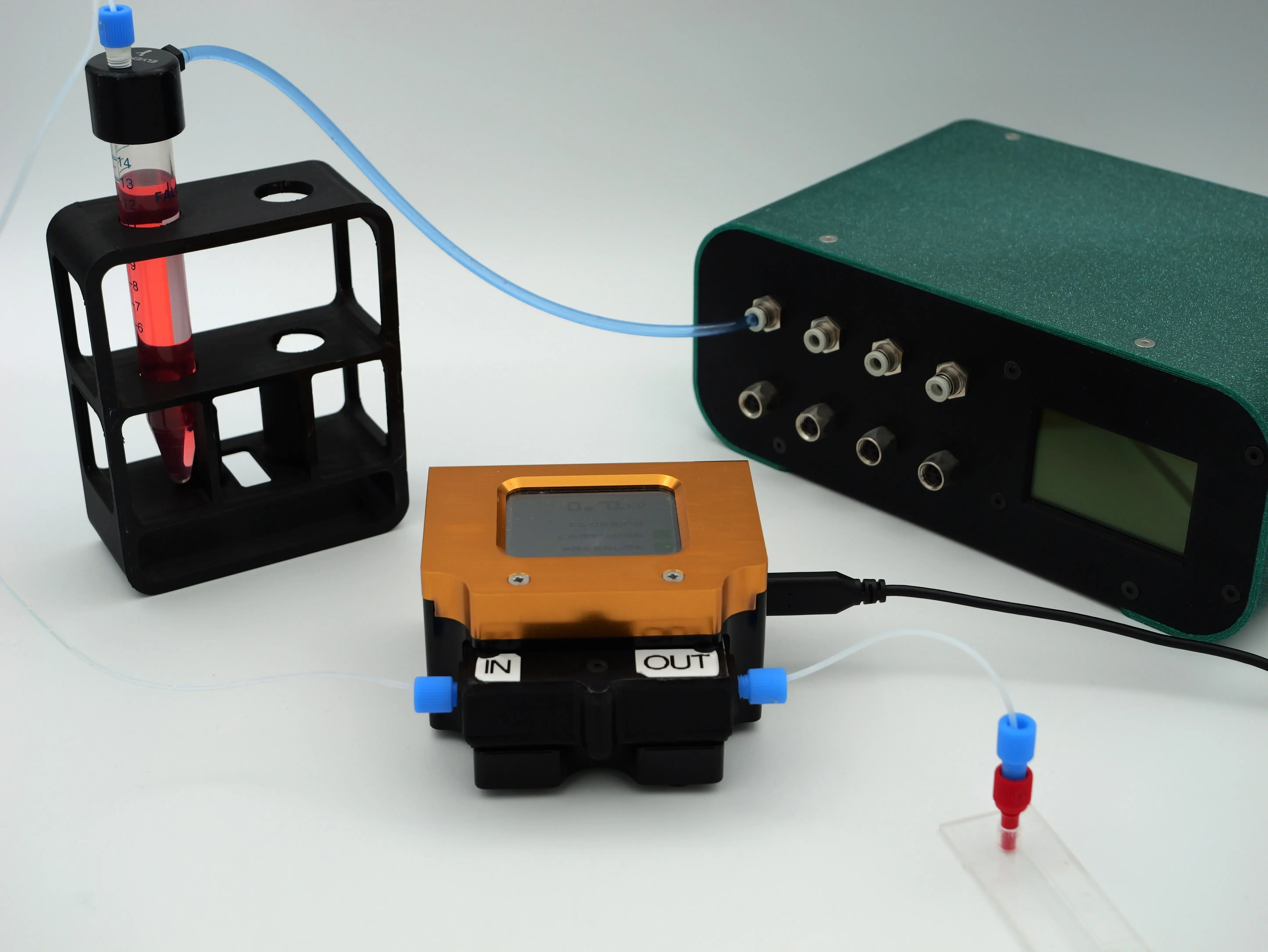
Inline viscometer unit
Single-use cartridges
Reservoirs, tubing and fittings
Dedicated software with CSV export
User guide
Spec sheet
Optional: flow sensor in the same line
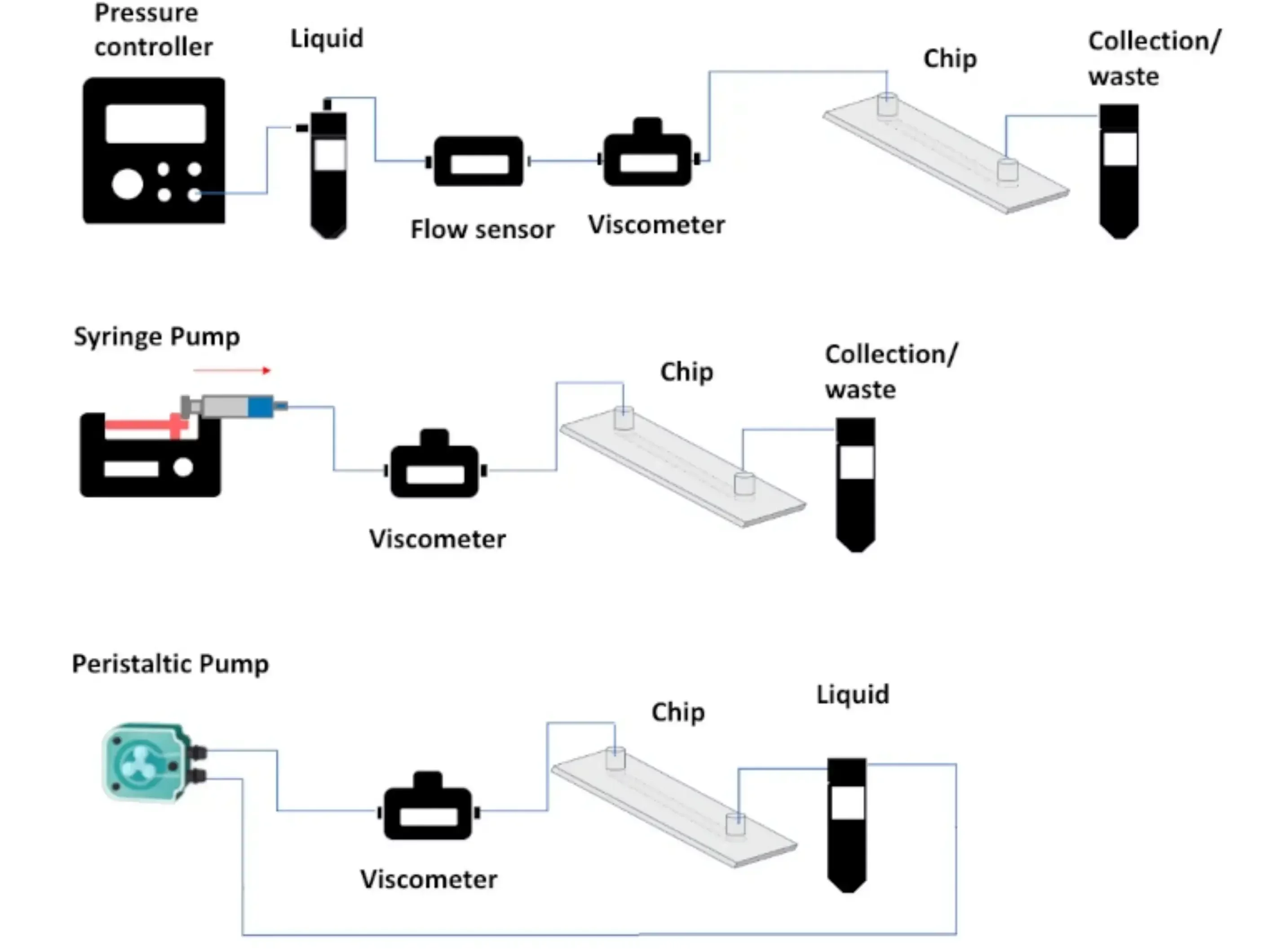
The viscometer runs with a pressure-driven flow controller, a syringe pump or a peristaltic pump. You can use it standalone, to characterise a medium before a run, or inline, where it monitors the perfusate through the culture. Used inline, it is set up so the shear used for characterisation stays away from the cells. The right arrangement depends on your loop, and that is a good reason to get in touch: send us your setup and we will scope how the viscometer sits in it.
Two practical points. Air bubbles disturb any inline microfluidic measurement, and a perfusion loop generates them, so degas the medium or fit a bubble trap upstream. And the wetted path is a single-use cartridge, supplied clean and sanitised, not supplied sterile. Tubing and connectors are available sterile.
Viscosity of cell culture media applications
Key applications include:
- Setting a target wall shear stress on endothelial or epithelial cells using the medium you actually perfuse, not water
- Tracking medium viscosity across a multi-day recirculating culture, so the shear stress reported on day three is the shear stress applied on day three
- Checking viscosity-modified media, with dextran or methylcellulose, against what the recipe predicted
- Comparing a serum-supplemented and a serum-free formulation, and knowing what changed hydrodynamically before blaming the biology
- Characterising each perfusate separately in a barrier model with two independent circuits, such as placenta, blood-brain barrier, gut or lung
- Supplying the viscosity correction for a flow sensor calibrated on water
- Reporting a measured viscosity in a methods section, so someone else can reproduce the shear stress
Barrier models with two recirculating circuits
MIC builds the microfluidic hardware behind organ-on-chip platforms, not the models themselves. In LIFESAVER, an H2020 Green Deal project on pharmacological risk assessment during pregnancy, MIC designed and built the barrier platform for a placenta-on-chip: independent media recirculation on the maternal and the foetal side, oxygen level measurement, and time-resolved sample collection on both sides [3]. Two media, recirculated independently over long runs, is precisely the case where the perfusate is neither water nor constant, and where a single assumed viscosity quietly propagates into every shear stress figure in the paper.
The same problem recurs in Bio-HhOST, an EIC Pathfinder Open project on 3D tissue models, where MIC develops a flow control platform for continuous, sterile, automated cell perfusion with live microscopy, without disconnecting the culture chip [4]. Longer runs mean more conditioning, and more reason to measure.
The inline viscometer was developed at MIC within GALILEO, an EIC Transition project, alongside a wide-range flow sensor and a flow calibrator [5]. It is offered for the applications above. It was not the instrument used in the projects that motivated it.
Inline viscometer technical specs
The inline viscometer comes with pre-calibrated liquids and the following specifications:
| Parameter | Value |
|---|---|
| Measurement | Shear-resolved viscosity, set by flow rate |
| Viscosity range | 0.2 to 100,000 cP |
| Shear rate range | About 1 to 1000 s-1 |
| Low-shear reach | Reliable down to 1 s-1 at organ-on-chip flow rates |
| Resolution | 0.01 cP |
| Accuracy | Better than 5% of reading |
| Repeatability | Better than 2% |
| Minimum sample volume | 0.1 mL, sample recoverable |
| Flow direction | Bi-directional, positive and negative flow |
| Operating temperature | 10 to 70 °C |
| Incubator compatibility | 37 °C, 5% CO2, up to 95% RH |
| Maximum operating pressure | 4 bar |
| Monitoring rate | Continuous logging up to 100 Hz |
| Configurations | Standalone, or inline in a perfusion line |
| Compatible pumps | Pressure-driven, syringe, peristaltic |
| Wetted path | Single-use PEEK and silicone cartridge |
| Cartridge | One per experiment, continuous runs up to 7 days |
| Sterilization | Not autoclavable, sanitization protocol available |
| Readout | On-device screen, software logging, CSV export |
| Connection | USB |
| Fittings | Standard microfluidic fittings, 1/4-28 or Luer |
| Chips | Commercial or home-made |
| Footprint | Compact benchtop unit |
Download the MIC Horizon Europe 2026/2027 Calls Calendar:
References
- [1] Šints V, Cīmurs J, Birjukovs M, Driķis I, Goluba K, Jēkabsons K, Parfejevs V, Riekstiņa U, Mozoļevskis G, Rimša R, Kitenbergs G. Physical model of serum supplemented medium flow in organ-on-a-chip systems. PLoS One. 2025;20(6):e0322069. doi:10.1371/journal.pone.0322069
- [2] Poon C. Measuring the density and viscosity of culture media for optimized computational fluid dynamics analysis of in vitro devices. J Mech Behav Biomed Mater. 2022;126:105024. doi:10.1016/j.jmbbm.2021.105024
- [3] LIFESAVER, Living Impact on Fetal Evolution: Shelter-Analyze-Validate-Empower Regulations. H2020, grant agreement no. 101036702. CORDIS
- [4] Bio-HhOST, Next Generation 3D Tissue Models: Bio-Hybrid Hierarchical Organoid-Synthetic Tissues Comprised of Live and Artificial Cells. HORIZON-EIC-2023-PATHFINDEROPEN-01, grant agreement no. 101130747. CORDIS
- [5] GALILEO, Innovative wide-range flow sensor to unlock microfluidic cell analyses. HORIZON-EIC-2022-TRANSITION-01, grant agreement no. 101113098. CORDIS






Funding and Support
This project has received funding from the European Union’s Horizon research and innovation program under HORIZON-EIC-2024-PATHFINDEROPEN-01, grant agreement no. 101185661(ERMES), HORIZON-EIC-2022-TRANSITION-01, grant agreement no. 101113098 (GALILEO). It has also received funding from the European Union’s Horizon research and innovation program under the Marie Skłodowska-Curie grant agreements no. 101119956 (DarChemDN).
About the author

Mukesh Kumar Sivakumar
Doctoral researcher in microfluidics, Microfluidics Innovation Center
Mukesh Kumar Sivakumar develops advanced microfluidic systems at the Microfluidics Innovation Center, as a doctoral candidate in the DarChemDN European doctoral network. He holds a BS-MS dual degree in physics from IISER Tirupati, India, and three years of hands-on research experience in microfluidics, biophysics, and diagnostics, in both academic and industrial settings.
ORCID | Google Scholar | ResearchGate | LinkedIn | DarChemDN
Products & Associated Accessories
FAQ: Inline viscosity of cell culture media
What does the inline viscometer do in an organ-on-chip setup, and what does it not do?
It measures the viscosity of the medium or perfusate flowing through it, in real time, at the temperature of the run, and it reports the shear rate each reading was taken at. That viscosity is the input a wall shear stress calculation, a CFD model or a shear stress calculator needs for your chip, with the rest coming from your channel geometry and your flow rate. Inside the measurement channel, shear stress is viscosity times shear rate, so it comes straight out of the same data. What the viscometer does not tell you is anything about cell viability, barrier integrity or medium composition. Those stay with your assays.
Is cell culture medium a Newtonian fluid?
Not always. Water is Newtonian. Serum-supplemented medium often is not. A 2025 study of DMEM with fetal bovine serum found shear-thinning behaviour, with a power-law flow index near 0.5 at 37 °C, and a wall shear stress an order of magnitude above the value calculated from the constant viscosity commonly quoted in the literature [1]. Earlier work found that a water model can still be adequate within a limited range of shear rates [2]. Whether the Newtonian assumption holds in your device depends on your geometry and your flow rate, which is why the medium is worth measuring rather than assuming.
How does the viscometer measure a shear-thinning medium?
The shear rate inside the measurement channel is set by the flow rate through it, so the instrument characterises the medium across a range of shear rates rather than at one point. The result is a viscosity curve for your medium, at the temperature you ran it. When the viscometer runs in a live culture line, the setup is arranged so the shear used for that characterisation does not reach the cells. The exact arrangement depends on your loop, and we will help you work it out.
Can the viscometer run inside a CO2 incubator at 37° C?
Yes. It is incubator-compatible and rated for 37 °C, 5% CO2 and up to 95% relative humidity, within a hardware operating range of 10 to 70 °C. This matters because viscosity depends strongly on temperature. A value read on the bench at 20° C is not the value the cells experience at 37° C.
How much medium does one measurement take, and do I get it back?
One measurement needs 0.1 mL. The sample is not consumed and can be collected downstream, in both the standalone and the inline configuration. That is the difference between characterising conditioned medium, a patient-derived sample or an expensive serum-free formulation, and deciding you cannot spare it.
Is the viscometer sterile, and can it run through a multi-day culture?
The wetted path is a single-use, disposable cartridge in PEEK and silicone, supplied clean and sanitised. One cartridge is intended for one experiment, with continuous runs up to seven days. It is not autoclavable and it is not supplied sterile. Tubing and connectors are available sterile. If your protocol needs a sterile wetted path for a multi-day culture, tell us what you need and we will scope the configuration with you.
Can I measure medium with dextran or methylcellulose added?
Yes. Raising medium viscosity with dextran or methylcellulose is a common way to reach a target shear stress at a lower flow rate. The viscometer measures what you actually made, rather than what the recipe predicted. Worth keeping in mind: an additive raises viscosity, but the shear response you get is the additive’s own, not a reproduction of any physiological fluid. Measuring across the shear rate range you intend to run is the only way to know the curve you have.
Does it work with peristaltic, syringe and pressure-driven pumps, and with reversing flow?
Yes to all three pump types. You connect its inlet to the outlet of your reservoir or syringe, and its outlet to your chip. Measurement is bi-directional, so positive and negative flow are both read, which covers withdrawal steps and setups that reverse the flow.
Can MIC join my Horizon Europe proposal as a microfluidic SME partner?
Yes. MIC is a French SME that has worked as the microfluidic engineering partner in H2020, Horizon Europe, EIC and MSCA projects, including the placenta barrier platform in LIFESAVER [3] and the automated cell perfusion platform in Bio-HhOST [4]. We take care of the microfluidic engineering, work on valorization, and optimize the proposal with you. If you are preparing a call and need a microfluidic SME partner, get in touch and let us know your needs.