Lung-on-a-Chip Model Pack
Perform lung research in a physiologically relevant microenvironment
Relevant microenvironment
Culture your lung cells in a physiological air-liquid interface
Up to 3-week long cell cultures
Continuous and controlled supply of nutrients in a stable flow
Fail-safe mechanism
Stop losing your cell experiment due to clogging
Use the chip of your preference
Choose the best parameters for your experiment

Need a microfluidic SME partner for your Horizon Europe project?
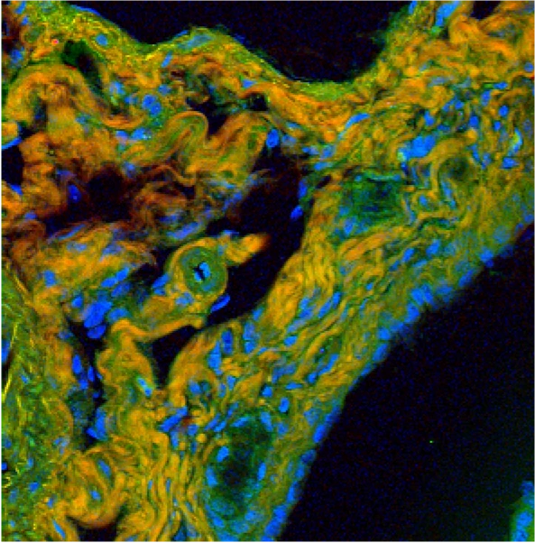
Image Caption: Ferret lung infected with canine distemper virus. Credit: Dr Stephen McQuaid & Stewart Church / QUB. Attribution 4.0 International (CC BY 4.0)
Why use a lung-on-a-chip model?
The limitations of current models for pathology research and drug discovery and screening are well-known. The standard culture of cells in static conditions has allowed the biomedical field to advance quickly, but it is a reductionist approach that cannot, by design, take into account most of the complexity in vivo [1].
Animal models do offer such complexity; however, the specificities of each species result in poor translation into humans and a huge loss of time and resources when moving from preclinical to clinical studies [2, 3].
The same is true in research focused on the respiratory tract. The lungs are one of the interfaces between the external environment and the body, being subjected to an air-liquid interface in a complex 3D structure, liquid flow, and constant mechanical stimulus.
These features are difficult to reproduce in in vitro models, as shown by Francis et al., in the table below [4]. The lung-on-a-chip model, based on the organ-on-chip technology derived from microfluidics, is the closest model to reproduce the in vivo setting in vitro without translational and ethical issues.
MultiDyn cell culture chamber
The Microfluidics Innovation Center partnered with IVTech, a biotechnology company specializing in advanced cell culture chambers to refine in vitro models.
MultiDynm, an evolution of the LiveBox1 chamber, has been designed and produced by IVTech SRL. The system is a rack of 4 wells in parallel, compatible in dimension with the column of a standard 24-well plate.
MultiDyn assures the opportunity to recreate all the experiments performed with LiveBox1, replicating the data in 4 parallel tests and reducing the complexity of the chamber setup. The rack is monitorable in real-time, using an inverted microscope, assuring the possibility of developing a 3D in-vitro model in dynamic conditions.
References
1. Mehling, Matthias, and Savaş Tay. “Microfluidic cell culture.” Current opinion in Biotechnology 25 (2014): 95-102.
2. Hoeck, Katrin. “How the Right In Vitro Biological Model Can Drive Success in Drug Discovery: Advanced physiologically relevant in vitro model systems can help predict the efficacy and safety of drug candidates early in the drug discovery process, reducing attrition rates.” Genetic Engineering & Biotechnology News 40.11 (2020): 19-21.
3. Van Norman, Gail A. “Limitations of animal studies for predicting toxicity in clinical trials: is it time to rethink our current approach?.” JACC: Basic to Translational Science 4.7 (2019): 845-854.
4. Francis, Isabella, et al. “Recent advances in lung-on-a-chip models.” Drug Discovery Today 27.9 (2022): 2593-2602.
Lung-on-a-chip pack setup
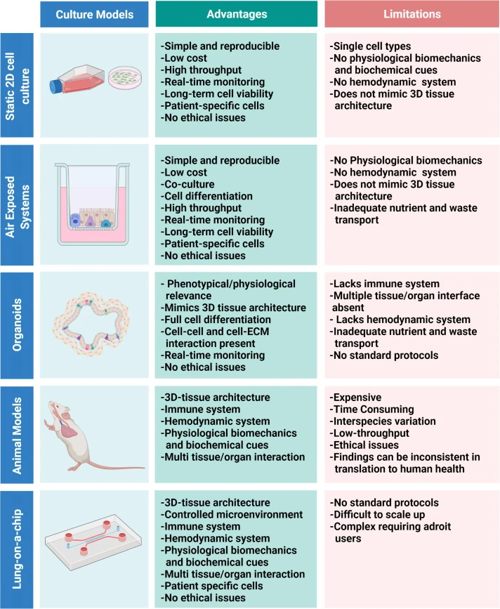
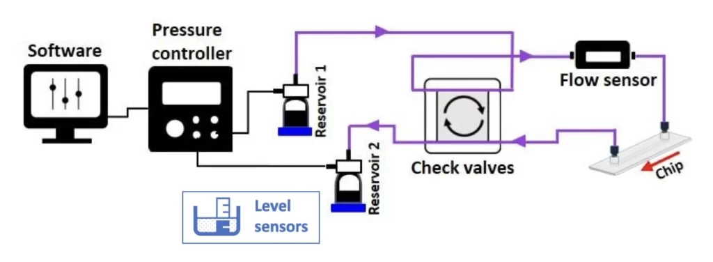
The lung-on-a-chip model can be perfused in a microfluidic chip for several days with a minimal volume of medium by using two media perfusion loops that can be integrated into our automated cell culture platform. To achieve that, this platform is based on a flow controller, a recirculation loop, flow sensors and level sensors.
With these additional features, the success of your microfluidic perfusion system is no longer dependent on the stability of your system’s fluidic resistance. In other words, you don’t need to worry about air entering your lines and damaging your cell culture due to fouling or clogging of the tubing!
In summary, the lung-on-a-chip model setup comprises the following instruments:
A typical lung-on-a-chip pack contains:
Flow sensor (Galileo, MIC)
Recirculation bridge
Level sensors
Software (Galileo user interface)
Flow controller
Several falcon reservoirs
Tubings and fittings
Microfluidic chip (Suggestion: ChipShop Fluidic 568)
User guide
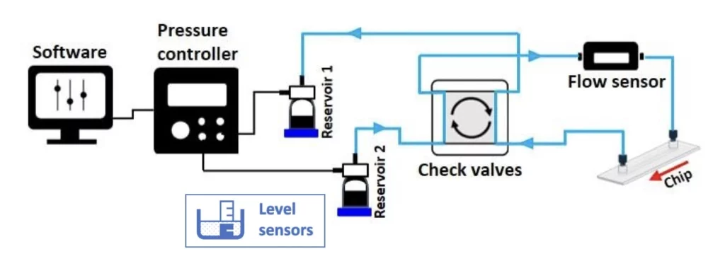
Lung-on-a-chip principle
The lungs are a barrier between the external and internal environments of the body, being characterized by an air-liquid interface. A lung-on-a-chip model reproduces this interface with a membrane chip, where one side of the membrane is flushed with air and the other with cell culture media or a blood-equivalent fluid. Each side of this membrane is laden with the appropriate cell types, and mechanical stimulus can be applied if the chip’s material is flexible.
Lung-on-a-chip applications
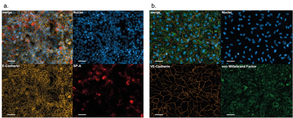
Deinhardt-Emmer et al. [2] developed a human alveolus-on-a-chip model using a membrane chip similar to the one suggested in this Pack. The goal was to develop a more physiologically-relevant model comprising endothelial and epithelial cells and macrophages to resemble the human alveolus architecture and functions.
The figure below shows each side of the membrane, using immunostaining to characterize each cell type. This same model was later used to study the inhibition effects of Pictilisib on influenza virus propagation [3] and to understand how the Sars-Cov-2 virus crosses the alveolar barrier to cause severe COVID-19 [4].
Check out our other organ-on-a-chip packs!
Gut-on-a-chip pack
Intestinal cells coculture under flow, mimicking the gut physiology
✓ All microfluidic pieces included, quick and easy assembly
✓ Dynamic culture conditions
✓ Advanced in viro/ex vivo
Gut-on-chip
Inflammatory bowel disease model
Automatically collect important markers of IBD in a relevant in vitro model
✓ Uncover cytokine profile changes in time
✓ Mimic pathological conditions of IBD
✓ Tailor sample volume to your analysis
Blood-brain Barrier on Chip
Plug-and-play instrument pack for long term BBB on a chip study
✓ Relevant microenvironment
✓ Automatized organ-on-chip perfusion
✓ Plug-and-play microfluidic platform
Blood-brain Barrier on Chip
Liver-on-a-chip model
Mimic the liver microenvironment in long term experiments
✓ Improve your reproducibility with physiological culturing conditions
✓ Automated and controlled supply of nutrients in a stable flow
✓ Test different conditions at the same time
Liver-on-chip
Bone-on-a-Chip Pack
Easily mimic the complex bone physiology and extracellular environment
✓ Control complex microenvironments
✓ Up to 3-week long cell cultures
✓ Fail-safe mechanism
✓ Plug-and-play platform
Bone-on-a-chip
Skin-on-a-chip
Reproduce the dynamic extracellular environment of the skin with ease
✓ Compatible with air-liquid interfaces
✓ Compatible with teer measurements
✓ Compatible with live cell imaging
Skin-on-a-chip
Kidney-on-a-chip
Physiological microenvironment for a more realistic in vitro kidney model
✓ Physiological flow rates
✓ Use the chip of your preference
✓ Enrich the media with metabolites
Kidney-on-a-chip
We have recently published a review of the different organ-on-a-chip models and current innovations.
References
1. Francis, Isabella, et al. “Recent advances in lung-on-a-chip models.” Drug Discovery Today 27.9 (2022): 2593-2602.
2. Deinhardt-Emmer, Stefanie, et al. “Co-infection with Staphylococcus aureus after primary influenza virus infection leads to damage of the endothelium in a human alveolus-on-a-chip model.” Biofabrication 12.2 (2020): 025012.
3. Deinhardt-Emmer, Stefanie, et al. “Inhibition of phosphatidylinositol 3-kinase by pictilisib blocks influenza virus propagation in cells and in lungs of infected mice.” Biomolecules 11.6 (2021): 808.
4. Deinhardt-Emmer, Stefanie, et al. “SARS-CoV-2 causes severe epithelial inflammation and barrier dysfunction.” Journal of virology 95.10 (2021): 10-1128.
Customize your pack
Our packs can be added to different setups depending on your specific needs. In this light, our microfluidic specialists will advise you on the best instruments and accessories depending on your needs and will accompany you during the system’s setup.
Frequently asked questions
How can we help your experiment?
This pack is in beta testing phase. So, although the instruments are not fully industrialized, we can provide extensive support as part of our beta testing program. Get in touch to see if you are eligible.
Can the pack be customized based on my sepcific application?
Yes! Our experts will establish which instruments are best suited for your application, such as the type of flow sensor or the number of flow controller channels you need to perform your experiment. Contact us using the “talk to our experts” green button above.
Can I buy individual instruments?
Our instruments are in beta testing phase and can be tested as a pack or individually, so get in contact with our team to know how our beta testing program works.
Download the MIC Horizon Europe 2026/2027 Calls Calendar:

Funding and Support
The ALTERNATIVE and LIFESAVER projects helped develop this instrument pack.
These projects are funded by the European Union’s H2020-LC-GD-2020-3, grant agreements No. 101037090 (ALTERNATIVE) and 101036702 (LIFESAVER).
Products & Associated Accessories
FAQ - Lung on a Chip Model
What is a lung-on-a-chip model?
A lung-on-a-chip is a microfluidic system that replicates the important physiological aspects of the human lung in vitro. It employs a membrane chip to replicate the interface of the air-liquid interface found in lung tissue where one side of the chip is exposed to the air and the other is continually perfused with cell culture media. Proper cell types are planted on both sides and mechanical stimulation can be used to imitate the breathing movement.
Why is a lung-on-a-chip model better as compared to traditional cell cultures or animal models?
The complexity of the in vivo environment of the lung including dynamic flow, mechanical stimulation, and the air-liquid interface can not be recreated in standard static cell cultures. Animal models are biologically complex and are poorly translated to other human physiologies, causing enormous loss of time and resources during preclinical-to-clinical translations. The lung-on-a-chip bridges both of these gaps, it offers physiological relevance, with neither the translational nor ethical constraints of animal testing.
What is included in lung-on-a-chip Pack?
A typical pack includes:
- A flow controller with fine accuracy.
- A flow sensor (Galileo, MIC)
- Check valves of a recirculation bridge.
- Level sensors
- Several falcon reservoirs
- Tubing and fittings
- Galileo user interface software.
- One microfluidic chip recommended (e.g., Chipshop Fluidic 568).
- A comprehensive user guide
What is the MultiDyn cell culture chamber? to what does it fit in this pack?
MultiDyn is an upgraded cell culture chamber invigorated by IVTech SRL, which is a collaborator of MIC that focuses on the refinement of in vitro models. It comprises a rack of 4 parallel wells that fits on a standard 24-well plate. MultiDyn allows simultaneous replicated experimentation of 4 parallel tests, real-time visualization of the experiment through an inverted microscope, and dynamic 3D in vitro culture conditions – it is a potent addition to the microfluidic perfusion setup.
What is the maximum time the cell cultures can stay in this system?
The pack will have the capability to sustain stable, continuous cell cultures to a maximum of 3 weeks. This is done by incorporating two media perfusion loops into automated cell culture platform that maintains a steady and constant supply of nutrients during the experiment.
What is the fail-safe mechanism and what is the reason?
Clogging or air penetration into the fluidic lines is a serious problem in microfluidic cell culture and may even annihilate long-term cell culture experiments. To prevent such occurrences, this pack includes level sensors and a recirculation bridge that consists of check valves that automatically detects and compensates that effect. Consequently, it is no longer the case that the success of the perfusion system is determined by the need to maintain uniform fluidic resistance – weeks of cell culture research are no longer at risk of failure.
What are the research applications of this pack?
Lungonachip model in particular suits these applications:
- Investigating alveolarcapillary barrier using endothelial and epithelial cocultures.
- Research on respiratory diseases (e.g., COPD, influenza, COVID19)
- Physiological screening of drugs and their effects.
- Understanding the mode of invasion of pathogens e.g. how SARSCoV2 enters the alveolar barrier.
- The pharmacological compounds are tested to determine those that act on lungspecific pathways.
Can the pack be customized for my experiment?
Yes. MIC specialists will check your application and suggest the most suitable setup, such as the type and number of flow sensor channels, the decision on microfluidic chip and any other modules it may need. It is also compatible with regular commercially sold chips of various brands making it even more flexible.
Should this pack be used by researchers who have no experience with microfluidics?
Yes. The pack is now in a beta testing stage, where MIC offers extensive practical assistance to users. The automated perfusion platform, combined with a single Galileo software interface, makes operating the system simple, and the user guide provides step-by-step instructions. Scientists are advised to reach out to MIC in order to identify their qualifications for the beta testing program.
Is it possible to buy separate instruments?
Yes, instruments can be tested and purchased separately or as a complete pack, depending on your laboratory’s requirements and available facilities. As the pack is in the beta testing phase, you can contact us to learn about the available options and how the beta program will be applied to access individual components.
