Live Cell Imaging
Keep ideal temperature and see your cells’ development in real time
Accurate temperature control
Keep your cells at 37°C ± 0.5°C outside the CO2 incubator
Up to 3 simultaneous devices
Test different conditions in the same system
For static or flow conditions
Designed for plates, dishes and perfused chips

Need a microfluidic SME partner for your Horizon Europe project?
Live cell imaging under perfusion
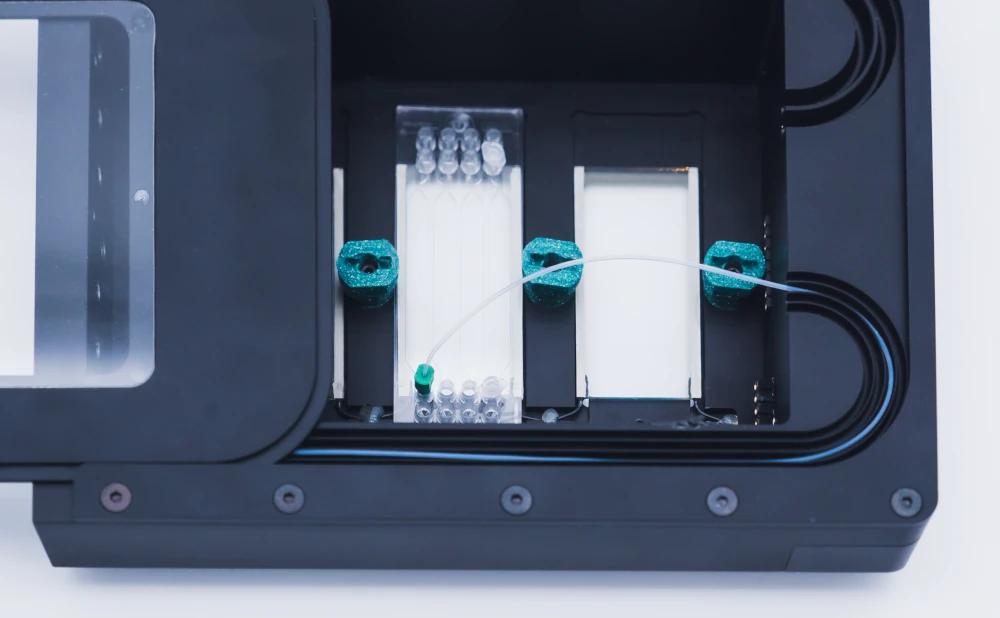
Our stage top incubator allows you to efficiently perform long-term live cell imaging experiments on top of the microscope stage. Designed to fit k-frame microscope stages, the internal chamber fits up to three parallel chips.
The ITO glass ensures homogeneous temperature across the microfluidic devices, and the frame was made so you don’t need to worry about heating your reservoirs. Your media will arrive at the right temperature on top of your cells regardless of whether you leave the reservoir at room temperature.
How does it work?
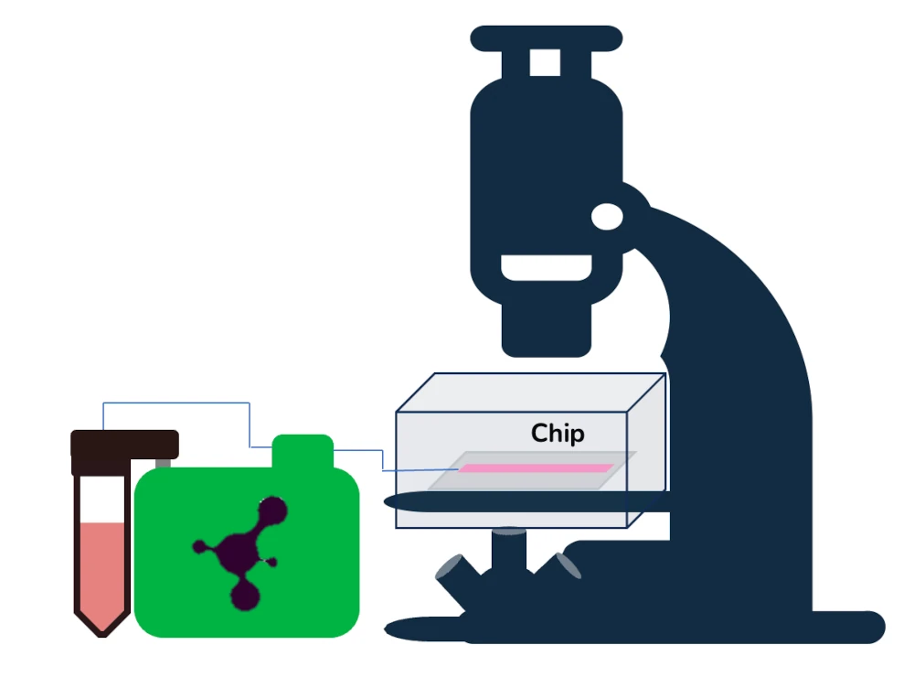
As a proof-of-concept of the suitability of the stage top incubator for live cell imaging, we cultured mouse microglial cells for 48 hours under continuous perfusion on top of the microscope stage. As shown below, the media flow control was done with a flow controller and a flow sensor.
Microglial cells, like other cell types from the brain parenchyma, are susceptible to shear stress as they are not usually exposed to flow in their physiological environment. Thus, to prevent the negative effects of shear stress, we employed the µ-Slide III 3D Perfusion and a microfluidic resistance to ensure a low and stable flow.
Live cell imaging pack setup
The live cell imaging pack includes:
Stage top incubator
Culture cells on top of the microscope stage for long periods
✓ Accurate temperature control
✓ Up to 3 simultaneous cell cultures
✓ High quality image gathering
Stage top incubator
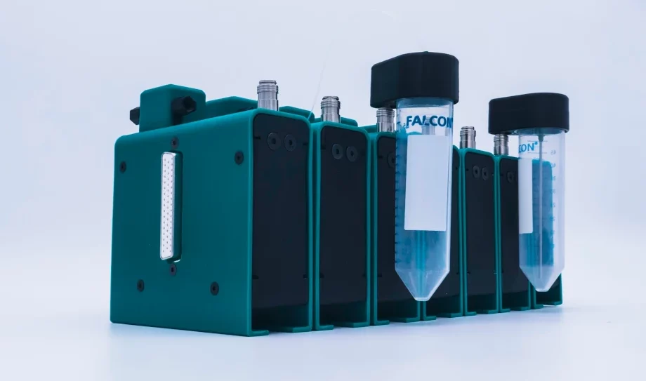
Perfusion pump for incubator
Culture up to 6 chips inside the incubator with the flow profile that you need
✓ Incubator-friendly
✓ No more mechanical stress
✓ Pulsatile or linear flow? You can choose
Perfusion pump
u-Slide III 3D Perfusion
Details of the experimental design can be found on the Specifications tab.
Live cell imaging specifications
Experimental details
According to the protocol published by Sepulveda et al., Glia, 2016, primary microglial cells were isolated from mice pups’ brains.
Primary mouse microglial cells were prepared and kindly provided by Patrick P. Michel and Rocio Gimenez from the Brain and Spine Institute in the frame of the LOCAI project.
After one week of isolation, cells were passaged onto ibidi chips (µ-Slide III 3D Perfusion, cat. 80376) at 150 000 cells/mL density. Cell medium was supplemented with FBS and PS. HEPES in powder was added to a final concentration of 20 mM. Once the HEPES dissolved, the pH was corrected and brought to 7.2 using Na 1 N, and then the media was filtered using a 0,22 filter. Cells were left to attach overnight in an incubator at 37°C and 5% CO2 before live cell imaging.
Technical specifications
Hardware
- Flow controller
- Wide-range flow sensor (Galileo)
- Tubings (1/32″ ID), fittings and reservoirs
- IBIDI µ-Slide III 3D perfusion
- 8 cm of 100 µm inner diameter microfluidic resistance
- Microfluidic male and female Luer to ¼” -28 connectors
- Laminar flow hood
- Standard Incubator for cell attachment, stage top incubator for experiment
Chemicals
- Dulbecco’s modified Eagle’s medium (DMEM)
- Penicillin/Streptomycin, 10,000 U/ml, 10mg/ml Streptomycin, PAN biotech
- FBS, 0.2 µm sterile filtered, PAN Biotech
- HEPES, to buffer pH in the absence of CO2 control.
Software
- Galileo user interface
- Thermal chamber software
Chip design
| Interface type | Female Luer |
| Well diameter | 5.5 mm |
| Well height | 1.7 mm |
| Volume per well | 30 μl |
| Material | Ibidi Polymer Coverslip |
| Surface treatment | ibiTreat |
| Growth area per well | 25 mm2 |
Live cell imaging results
Live cell imaging allows for studying the dynamic aspect of cellular processes. In vitro models, however, need to be under strict environmental control. Parameters such as pH, temperature, and oxygen levels are critical for robust and reliable results. Whether the experimental paradigm requires shear stress, continuous perfusion, or static conditions, the need for accurate temperature regulation is the same.
The videos below show 8h time-lapses of primary microglial cells taken immediately after lipopolysaccharide (LPS) stimulation. These videos were recorded using microglial cells seeded onto microfluidic chips and either left under static, non-perfused conditions (video 1) or connected to a pumping system for continuous perfusion along the experiment (1 µL/min, video 2).
Video 1. Microglial cells were seeded in a µ-Slide III 3D Perfusion chip well and maintained in the stage-top incubator on the microscope frame in static conditions for 8 hours after LPS stimulation. Cells were challenged with 10 ng/mL LPS at t = 0 h. Micrographs in phase contrast were acquired every 30 minutes for 8 hours. Image acquisition was made using an AxioVert inverted microscope with a 20x magnification objective (Zeiss).
Video 2. Microglial cells were seeded in a µ-Slide III 3D Perfusion chip well and maintained in the stage-top incubator with continuous perfusion at a 1 µL/min flow rate. Cells were challenged with 10 ng/mL LPS at t = 0 h. Micrographs in phase contrast were acquired every 20 minutes for 8 hours. Image acquisition was made using an AxioVert inverted microscope with a 20x magnification objective (Zeiss).
In both cases, the incubator allowed for the recording of microglial cells. Micrographs taken every 20 or 30 minutes show these cells’ highly dynamic and motile nature. Significantly, microglial cells, like other cell types from the brain parenchyma, are not exposed to mechanical stresses under physiological conditions. They require nutrient delivery and removal of waste metabolites for survival as in physiological conditions.
The stage top incubator presented here allows for flexibility in designing experiments, including when deciding to apply specific mechanical stress linked to flow. Several off-the-shelf chips with different geometries will allow us to extend the design possibilities to address physiological or pathophysiological conditions using the tissue or cell type of choice.
Customize your pack
Our Packs can be modified depending on your specific needs. Our microfluidic specialists will advise you to provide the best instruments and accessories, depending on your needs. They will accompany you during the setup of the microfluidic platform.
Frequently asked questions
What are the dimensiosn of a K-frame stage top?
For all the specifications of the stage top incubator and its compatibilities, please check the dedicated page here.
Can I order a pack?
Our Packs, such as the Live Cell Imaging pack, are available under specific conditions. As these packs are still in the developmental stage, we have some eligibility criteria to ensure their optimal success rate.
At the Microfluidics Innovation Center, we tailor our approach to suit each researcher’s unique needs. Therefore, we encourage you to contact our technical team for a detailed discussion of your research objectives and requirements.
To learn more about our Packs or to schedule a consultation with our experts, please contact us using the “talk to our experts” green button above. We look forward to collaborating with you and supporting your research endeavors.
Can I buy individual instruments?
Our instruments are in beta testing phase and can be tested as a pack or individually, so get in contact with our team to know how our beta testing program works.
Is the stage top incubator gas-tight?
No, the Stage Top Incubator allows gas exchange with the atmosphere.


Funding and Support
The ACDC, and LOCAI projects results helped develop this pack, with funding from
the European Union’s Horizon 2020 research and innovation program under grant agreement No 824060 (ACDC project),
and French National Research Agency (ANR) and the German Federal Ministry of Education and Research (BMBF) in the frame of the 1st German-French Joint Call for proposals on Artificial Intelligence.
Products & Associated Accessories
FAQ - Live cell imaging
What is the problem that this Live Cell Imaging pack is actually solving?
It is designed with a highly narrow pain point of long-term microscopy on living cells without compromising environmental controls the moment you remove your culture out of the CO2 incubator. The pack is aimed at maintaining temperature on the microscope stage (within physiological range) to allow you to monitor cell dynamics in real time rather than undergoing short grab and run imaging sessions.
Do microfluidic chips only, or can plates and dishes also be used?
It is designed on plates, dishes, and microfluidic chips that have performed perfusion. The main principle is simple, keep your sample hot photo constantly. Suppose today you have a plate based arrangement but you want to move to the perfusion chips (or the other way round), the pack is supposed to remain handy instead of compelling you to adopt a single inflexible format.
4)What is the number of experiments that I can perform simultaneously on the microscope?
The chamber at the top of the stage has a size that fits up to three parallel devices/chips on a k-frame microscope stage. The same thing about that 3-in-parallel nature: not only is it convenient that way, it allows you to juxtapose conditions (e.g. static vs perfused, drug A vs drug B, or two flow regimes plus a control) with less day-to-day variability.
What about the ITO glass you refer to it is not that important anyway?
Yes, and not in a marketing way. The incubator will be made of ITO glass to contribute to the even distribution of heat over the devices. Equalization of heating across the chip footprint means that local gradients are reduced, so that is one of the micro-level problems that certainly manifests as edge effects, non-uniform migration velocity, or non-reliable phenotype changes.
Is this able to perform the static culture and perfusion?
Exactly. The pack data given in demonstration was in the form of non-perfused conditions (static) and continuous perfusion. An example is with a constant perfusion of 1 uL/min when imaging. That is a reasonable voltage spread of a lot of delicate primary cells when you need to replenish nutrient in the cell without taking undue mechanical abuse.
What does the pack contain?
At least, the presented setup is a combination of:
-A stage-top on-microscope long run (up to 3 parallel) incubator.
-A flow control unit and a broad-range flow sensor (closely coupled with a special user interface).
-Tubing and fittings (especially 1/32-inch inner-diameter tubing), reservoirs, and standard microfluidic connectors (including Luer-to-microfluidic thread adapters).
-The microfluidic resistance element was 8 cm long, with a 100 µm inner diameter, to stabilize/limit flow.
-A perfusion pump for use in the incubator, capable of growing up to 6 chips, with the option of pulsatile or linear flow profiles (useful when resting to recreate vascular conditions or when you need to avoid the pump’s effects on a cell line), is also mentioned.
Does the stage-top incubator have a gas-tight? What does that mean in the way to control pH and oxygen?
It is clearly stated that it is not gas-tight in nature, i.e. it exchanges gas with the atmosphere. That is not necessarily bad either, it just implies that you usually regulate pH differently than in a closed incubator of CO2 (which is why HEPES buffering was used in the given protocol-style description). In case your experiments are highly CO2 sensitive or demand a specific hypoxia/hyperoxia, you will need to consider the gas strategy (buffering, formulation of media, and any upstream gas conditioning).
What is the rationale behind incessant talk about shear stress – is it a pack of fragile cells?
The use case is biased in favor of cells which do not inherently undergo a flow condition, the shear can be a confounder. A good case in point is microglia: they are said to be especially vulnerable to shear, in the sense that parenchyma in the brain is not a flow-exposed space as in the case of endothelium. The system solves this by using a combination of controlled perfusion (combined with flow damping (resistance + stable control) in order to achieve low, steady flow instead of unintentional pulses or spikes.
Is it a standard product or is it still experimental?
It is being offered as a pack under certain conditions, and the eligibility requirements are due to the fact that it is a product at a developmental / beta-stage. Practically, that normally represents: they will desire to realize your microscope limitations, chip format, biological limitations (shear sensitivity, sterility demands), and what success will appear like in your undertaking to the point where they will commit the precise configuration.
