Bio-HhOST
Cell interaction in next generation 3D tissue models
Writer
Celeste Chidiac, PhD
Keywords
Microfluidic Devices, Intelligent Microfluidics, Artificial Intelligence, Machine Learning
Author
Marlene Kopf
Publication Date
June 24, 2024
Keywords
Intelligent Microfluidics
Deep Learning
Microfluidic Devices
Artificial Intelligence
Machine Learning
3D tissue models
Cell interaction
Cell perfusion

Your microfluidic SME partner for Horizon Europe
We take care of microfluidic engineering, work on valorization and optimize the proposal with you
An interdisciplinary consortium to observe cell interaction
The Bio-HhOST project aims to develop bio-hybrid materials integrating living and artificial cells to observe cell interaction, allowing artificial cells to influence the growth, differentiation, and function of living cells. We are working on Bio-HhOST with interdisciplinary team of biologists, engineers, mathematicians, and entrepreneurs.
In the project Bio-HhOST, we aim to create precision-engineered artificial cells made of liquid and lipid bilayers, compartmentalized chemically, and co-located with live cells to induce cell interaction. These artificial cells will have functional metabolisms and respond to environmental chemical stimuli by releasing signaling molecules to regulate neighboring living cells, mimicking complex biological tissues and their cell interaction.
Project goal: understanding cell behavior in realistic 3D tissue models
The overarching goals of the project include the creation of next generation 3D tissues. In these tissues, living cell differentiation is spatially regulated by chemically programmable artificial cells. The 3D tissues are intended to be maintained through cell interaction and dynamic communication between live and artificial cells. In addition, the project partners will develop multi-level models of organoid-synthetic tissue behavior and cell interaction for targeted applications. Another important part of the project deals with the evaluation of drug delivery vectors for next-generation biological therapeutics. Moreover, complex tissues with distinct regions that are not achievable with current organoid protocols will be generated in the scope of the project.
The broader ambitions of Bio-HhOST include understanding cell behavior in realistic 3D tissue models to observe cell interaction, identifying disease treatment targets, and reducing the use of animals in pharmaceutical research. With this innovative approach, the project aims to enhance the understanding and control of physio-chemical cell interaction in tissues, offering significant advancements in biomedical science and therapeutics.
How to use microfluidics for next generation 3D tissue models: our role
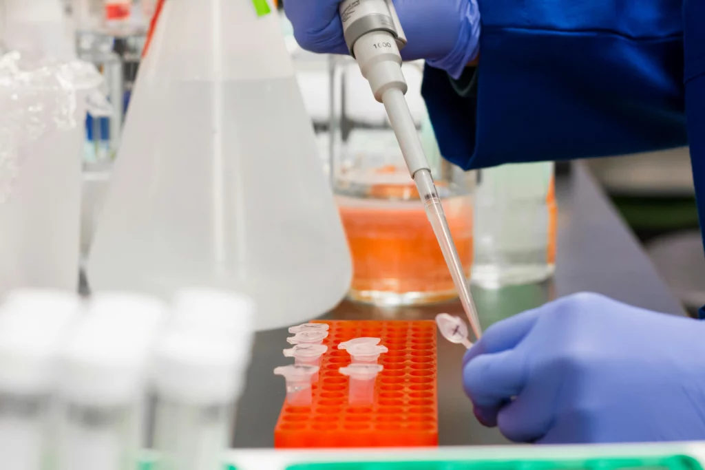
 The Microfluidics Innovation Center is working on a microfluidic flow control platform for automated cell perfusion for the Bio-Hhost project. The platform enables researchers to regulate cell growth parameters and observe cell interaction without the need for an incubator. The system will allow continuous manipulation under sterile conditions and ongoing monitoring via microscopy without disconnecting the cell culture chip from the perfusion setup.
We have also been involved in other projects related to artificial cells and cell interaction. Find out more about the projects ACDC and Protomet on our website. More information on ACDC can be found on the project’s website.
You need a microfluidics partner for your project on artificial cells as well? Drop us a line! We are interested in joining your consortium and are open to a wide variety of topics and calls, such as the Pathfinder Challenge on Solar-to-X devices dealing with biohybrid systems.

Funding and Support
This project has received funding from the European Union’s Horizon research and innovation program under HORIZON-EIC-2023-PATHFINDEROPEN-01, grant agreement no. 101130747 (Bio-HhOST).
Start date: 1 February 2024
End date: 31 January 2027
Overall budget: € 1,226,718.83
Check our Projects
FAQ – Cell interaction in next generation 3D tissue models - Bio-HhOST
In a nutshell, what is Bio-Hhost attempting to construct?
A mini-tissue that is controllable, where living cells and programmable artificial cells communicate with one another. Artificial cells are liquid compartments enclosed in lipid bilayers that detect cues in their microenvironment and emit signals to direct the growth, differentiation, and function of surrounding living cells. Imagine organoid biology with an added step of chemical decision-making.
What is the importance of this to 3D tissue models?
The majority of organoids lack the refined communication mechanisms of real tissues, including spatially resolved signals, gradients, and cell-cell communication. Bio-HhOST, by incorporating artificial cells into its design that can manufacture or detect signals on demand, aims to rediscover such physiology, so polarization, barrier formation, immune synapses, and stromal cross-talk will become more robust and extend culture times.
What are the artificially-generated cells here?
Lipid-bilayer interface chemically compartmentalized droplets. They can support basic metabolic processes, respond to environmental cues (such as chemical signals), and communicate in a timely or controlled manner. They are co-localized with living cells and act as programmable neighbors, pushing the tissue in the way it desires.
What does MIC have under construction on the project?
An automated and sterile cell perfusion microfluidic flow-control platform designed to allow continuous culture maintenance and imaging of cultures without removing chips in and out of an incubator. The apparatus maintains temperature, flow, and nutrient exchange, and holds the chip in place under a microscope during actual long-term experiments.
What fraction of microfluidics biology occurs in comparison with biology on static gels?
Under flow, you can provide morphogens as graded profiles, eliminate waste before it concentrates, and have separate “neighborhoods” (epithelium, stroma, endothelium, immune cells) with independent perfusion loops. What is obtained is less fluctuating phenotypes and interaction patterns that can be maintained longer than a week, right where most stagnant cultures wander.
Concisely, what are the scientific objectives of the project?
Three pillars: (i) engineer next-gen 3D tissues in which differentiation can be spatially programmed with chemically programmable artificial cells; (ii) simulate multi-scale organoid-synthetic tissue behavior; and (iii) screen drug-delivery vectors and form complex tissues with spatially localized differentiation that protocols based on standard organoid procedures cannot.
Readout strategy, more than pretty pictures?
Function pairs with imaging. Aim to capture interaction-mediated transcriptional programs. Expect barrier integrity (TEER and tracer permeability) under flow, live imaging of immune contacts and cytokine bursts, secretome measurements, and single-cell omics. Where feasible, data are compared to ex vivo tissue slices or primary samples.
Is this automation and screening compatible?
Yes. The platform is geared towards robotic liquid handling, environmental control, and multiplexed readouts (imaging, ELISA-style secretome panels, qPCR/NGS). MIC is the bread-and-butter of enabling lab protocols to be turned into SOP-based, standardized connector and plate-form footprints, thereby improving throughput.
What is different in drug discovery or advanced testing?
Now you can probe therapies whose functionality relies on cell-cell communication-biologics, cell therapies, vectorized payloads, within micro-architectures conformed to the target tissue in terms of gradient, stiffness, and spatial organization. In practice, a team begins with a focused assay, which is scaled to semi-throughput once the biology is stable.
We are building a Horizon Europe consortium- what is the need to include MIC?
Since an experienced SME risks away engineering and expedites exploitation plans, MIC has co-written work packages, harmonized on-site SOPs, and transformed prototypes into powerful tools across various EU projects. Our portfolio consortia that incorporate MIC generally achieve a success rate of about 2x that of the official first-submission baseline, but this is by no means a guarantee, and this trend is repeated with high frequency when prototyping and valorization are taken into account from the very beginning.
What are the requirements of partners to initiate a joint work package?
A no-nonsense biological query (which interaction and what success look like), access to primary cells or well-defined lines, and the needed readouts. We will do micro-engineering, automation, and validation; we will also format proposals, milestones, risks, and IP/exploitation in a way that allows the impact to be tracked.
