Speroid Cell Culture Microfluidic Platform
Plug-and-play pack for automated scaffold-free 3D cell culture technique
Multiple parallel culture of spheroids
Depending on the chip, more than 20 spheroid can be cultivated
More physiologically relevant model
Dynamic perfusion better mimics real conditions than static culture
Automated spheroid perfusion
Perfuse spheroids for several days with automated sequences

Need a microfluidic SME partner for your Horizon Europe project?
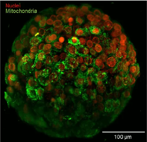
Breast cancer cell spheroid, SEM picture Dr Khuloud T. Al-Jamal (CC BY 4.0)
A microfluidic beginner instrument pack for spheroid cell culture
Spheroids are sphere-shaped cell cultures in a 3D scaffold that let cells proliferate and migrate inside the scaffold to reproduce cell configuration inside the human body. Drugs can be tested using this model without animals and with better mimicking of cell morphology, physiology, and organization compared with 2D cell culture inside Petri dishes.
Spheroids can be perfused with microfluidic instruments to obtain better reproducibility, continuous physiological shear stress, long automated culture experiments, use of costly fluids, and better control of parameters like pH or temperature, making it the most efficient method to culture spheroids.
We assembled a beginner-friendly pack with high-precision microfluidic instruments, including a flow controller with flow sensors for continuous flow rate control that you can combine with your homemade microfluidic chip or a commercially available one.
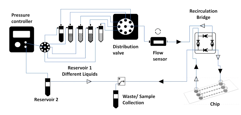
Microfluidic spheroid cell culture setup
We assembled a microfluidic platform to perfuse and induce spheroid growth with perfectly well-controlled parameters. This plug-and-play pack is perfectly suited for researchers who want to transition from batch static culture to microfluidic dynamic cell culture for single spheroid observation, for example.
This beginner-friendly microfluidic pack for spheroid culture, observation, and screening is combined with the flexible Elveflow software for sequence setting and automation, allowing improved reproducibility and sample use optimization.
An accurate flow controller provides a totally pulseless and fast response flow control coupled with flow rate sensors (Galileo). Pressure-driven systems are the most stable and precise microfluidic solutions on the market, especially in comparison to syringes and peristaltic pumps. This is especially important to perfectly control the applied shear stress on the cells to better mimic in vivo conditions.
For long-term experiments like spheroid culture in microfluidic chips, recirculation of the medium is needed to maintain a constant, sufficient shear stress applied to the cells without using large quantities of expensive medium. Several methods exist to perform recirculation; a recirculation bridge or active valves can be used.
A sequential injection valve can also be added to inject different media and drugs. A commercialized dedicated microfluidic chip for spheroids can also be included.
The spheroid cell culture pack is highly customizable and can include several instruments to form the following platform. Each instrument is compatible with the others, piloted by the same software, and has a dedicated user guide for beginner-friendly step-by-step setup.
The spheroid cell culture pack contains:
Flow sensor (Galileo, MIC)
Software (Galileo user interface)
Recirculation bridge
Sequential injection valve
Flow controller
Fittings, tubings & luers
Reservoirs
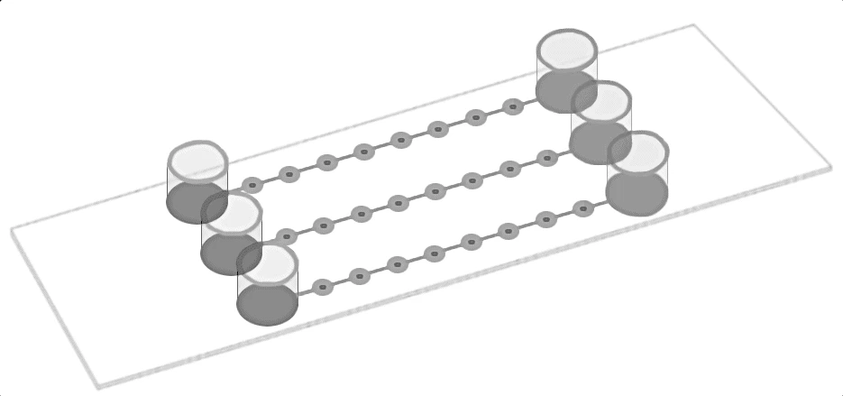
Microfluidic chip for spheroid cell culture
User guides for instruments
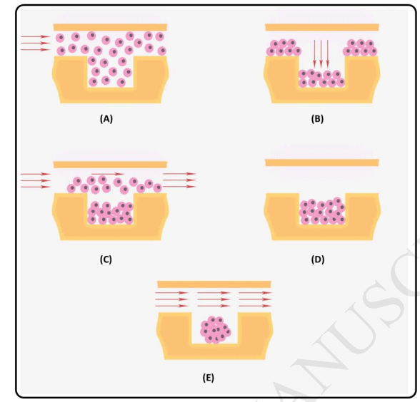
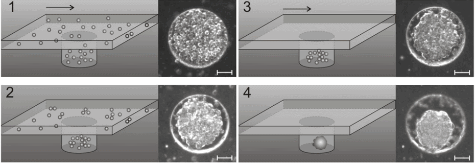
Spheroid microfluidic cell culture principle
Spheroid, as three-dimensional cell structure, better reproduce cell-to-cell interactions than monolayer cultured cells and generally mimick in vivo environment more closely [1]. Spheroids are considered to be the best tumor cellular models [2] but can also be used as models for neurodegenerative diseases [3].
It has been shown that using microfluidics for spheroid cell culture is a great tool to test drugs with better accuracy, throughput, and microphysiological in vitro tests while reducing the need for animal models [5].
The other advantages include the possibility to culture spheroids with different sizes, decrease experiment costs and energy consumption, continuous and controlled physiological shear stress, and the possibility to observe a single spheroid at a time [6-7]. Microfluidic spheroid platforms have been successfully used for drug screening using different chip designs [8-10].
References
Astashkina, Anna, Brenda Mann, and David W. Grainger. “A critical evaluation of in vitro cell culture models for high-throughput drug screening and toxicity.” Pharmacology & therapeutics 134.1 (2012): 82-106.
Friedrich, Juergen, Reinhard Ebner, and Leoni A. Kunz-Schughart. “Experimental anti-tumor therapy in 3-D: spheroids–old hat or new challenge?.” International journal of radiation biology 83.11-12 (2007): 849-871.
Słońska, Anna, and Joanna Cymerys. “Application of three-dimensional neuronal cell cultures in the studies of mechanisms of neurodegenerative diseases.” Postepy Higieny i Medycyny Doswiadczalnej (Online) 71 (2017): 510-519.
Moshksayan, Khashayar, et al. “Spheroids-on-a-chip: Recent advances and design considerations in microfluidic platforms for spheroid formation and culture.” Sensors and Actuators B: Chemical 263 (2018): 151-176.
Petreus, T., Cadogan, E., Hughes, G. et al. Tumour-on-chip microfluidic platform for assessment of drug pharmacokinetics and treatment response. Commun Biol 4, 1001 (2021).
Kim, Jong Bin. “Three-dimensional tissue culture models in cancer biology.” Seminars in cancer biology. Vol. 15. No. 5. Academic Press, 2005.
Karina Ziółkowska; Agnieszka Stelmachowska; Radosław Kwapiszewski; Michał Chudy; Artur Dybko; Zbigniew Brzózka (2013). Long-term three-dimensional cell culture and anticancer drug activity evaluation in a microfluidic chip. , 40(1),
Kwapiszewska, K., et al. “A microfluidic-based platform for tumour spheroid cell culture, monitoring and drug screening.” Lab on a Chip 14.12 (2014): 2096-2104.
Lim, Wanyoung, and Sungsu Park. “A microfluidic spheroid culture device with a concentration gradient generator for high-throughput screening of drug efficacy.” Molecules 23.12 (2018): 3355.
Patra, Bishnubrata, et al. “Drug testing and flow cytometry analysis on a large number of uniform sized tumor spheroids using a microfluidic device.” Scientific reports 6.1 (2016): 1-12.
Pampaloni, Francesco, Nariman Ansari, and Ernst HK Stelzer. “High-resolution deep imaging of live cellular spheroids with light-sheet-based fluorescence microscopy.” Cell and tissue research 352.1 (2013): 161-177.
Why use microfluidics for spheroid cell culture?
Culturing spheroids in microfluidics brings critical advantages in comparison to classical methods:
- Decrease experiment costs by reducing the amount of reagent.
- Physiological shear stress is applied to the spheroid.
- A single spheroid can be cultivated and observed.
- Even long experiments can be easily automated and don’t require human intervention.
- Improved oxygen and nutrition supply for cells.
- Better reproducibility and uniformity.
- Easily inject precise volumes of different drugs or compounds.
These advantages make microfluidics the best solution to perform spheroid culture and drug screening. Furthermore, our instruments are especially well suited for this application because they are stable, user-friendly, and accurate and provide the best flow control on the market overall.
References
Karina Ziółkowska; Agnieszka Stelmachowska; Radosław Kwapiszewski; Michał Chudy; Artur Dybko; Zbigniew Brzózka (2013). Long-term three-dimensional cell culture and anticancer drug activity evaluation in a microfluidic chip, 40(1).
Customize your spheroid cell culture pack
Several commercialized and laboratory made microfluidic chips have been developed and tested to perform spheroid cell culture. We can include one of them with different surface modifications.
This pack can be customized depending on your specific needs. Microfluidic chips are transparent and can be easily combined with microscopy to observe the growth of the spheroids.
This pack can be personalized to cultivate different sorts of spheroids. Our experts will help you determine your needs depending on your application and provide continuous and full customer support to fulfill your experiment goals.
Contact our experts to answer any questions about this spheroid cell culture pack and how it can match your specifications!
Download the MIC Horizon Europe 2026/2027 Calls Calendar:

Funding and Support
The Orgtherapy project results helped develop this instrument pack; it has received funding from the French Agence Nationale de la Recherche (ANR) in the frame of ERA-NET JPco-fuND 2019.
Products & Associated Accessories
FAQ - Spheroid Cell Culture Pack
What is a spheroid, and why do we use it in cell culture research?
A spheroid is a sphere-shaped, three-dimensional cell aggregate that allows cells to proliferate and migrate in a way that closely mimics their natural organization inside the human body. Unlike flat, monolayer cultures in Petri dishes, spheroids replicate cell-to-cell interactions and the physiological architecture of living tissue. They are considered the gold standard tumor cellular model, but are also widely used to study neurodegenerative diseases and test drug efficacy without relying on animal models.
What are the drawbacks of spheroid traditional monoculture?
Normal batch static culture does not provide dynamic conditions as provided in vivo. It is poorly reproducible, has little control over factors such as pH, temperature, and shear stress, and automation of long-term experiments is difficult. The rate of reagent use is high as well, and there is virtually no possibility of isolating and studying just one spheroid over time. These limitations reduce the validity of drug screening findings and increase the cost of experiments.
What is the benefit of microfluidics in enhancing the culture of the spheroid cell?
Microfluidic perfusion presents continuous, regulated fluid flow through the chip, and this presents a number of tangible benefits:
- Physiological shear stress is used incessantly, making it more in vivolike
- Nutrient and oxygen supply to the cells is enhanced
- Longterm experiments can be automated and do not require human involvement
- Strict doses of drugs or compounds can be injected in sequence
- There is a high reduction in the reagent and medium consumption
Differentiable and consistent reproducibility between spheroids is improved.
What is the MIC Spheroid Cell Culture Pack?
The pack is a plugandplay platform that enables the transition from static to dynamic microfluidic culture for researchers. It includes:
- A pressuredriven flow controller without pulses,
- Flow sensors for continuous flow rate monitoring (Galileo, MIC),
- A recirculation bridge to sustain constant shear stress.
- A sequential injection system to inject various media or drug substances,
- Fittings, tubings, reservoirs, and an optional dedicated spheroid chip are all compatible.
- Automation software and control of the parameters of sequences,
- User instructions on a stepbystep basis for each instrument.
All the parts are compatible and operate through the same software interface.
What advantages does pressure-driven flow control over syringe or peristaltic pumps have on the culture of the spheroid?
Pressure-driven systems generate stable, highly responsive flow. This is extremely important in spheroid culture, since any change in flow is directly converted into an uneven shear stress on the cells. Syringe pumps cause mechanical pulsations, and peristaltic pumps cause periodic pressure fluctuations, both of which undermine the physiological relevance of the culture model. The flow controller in the MIC pack offers better accuracy and stability, which are necessary for long, controlled experiments.
Does long-term medium recirculation play a role in long-term spheroid experiments?
Continuous replenishment of fresh medium in a single-pass arrangement is prohibitively expensive, particularly with special or expensive media, in experiments that require more than a few days. Recirculation enables the same medium to be recirculated through the chip several times using a recirculation bridge or active valves, ensuring that shear stress remains constant and sufficient nutrients are delivered without requiring large volumes. This contributes to the economic feasibility and the strength of the long-spheroid culture experiments.
In which applications is this platform applicable?
The spheroid cell culture pack is applicable to:
- The development of tumor models and screening of oncology drugs,
- A 3D modeling of neurodegenerative disease with neuronal spheroids,
- Pharmacokinetic and drug efficacy evaluation under physiological flow,
- Highthroughput screening using multispheroid chip configurations (up to 20+ spheroids in parallel).
- Single spheroid observation and longitudinal monitoring under a microscope.
The transparent chip design also allows them to be directly integrated with optical and fluorescence microscopy.
How user-friendly is the pack to particular research requirements?
It has a very modular platform. It can be modified by researchers to suit a variety of different chip geometries and surface modifications, flow configurations, or by adding other injection valves to support multi-drug protocols. MIC experts are offering special assistance to align the platform with cell types, sphere sizes, and experimental endpoints. Any commercially available chip and the design made in the labs can be used with the system.
How can MIC be involved in a Horizon Europe consortium?
MIC is a microfluidics SME with a long history of involvement in EUfunded research consortia, including projects sponsored by the French ANR within ERANET frameworks. In a Horizon Europe project, MIC can play a role as a nonacademic beneficiary or technology partner, which can offer:
- Microfluidic hardware and automation parts of complicated bioassays,
- First deliverables, which are prototypical and anchor work packages with real, tangible milestones,
- Development of applicationspecific platforms, including organonchip and diseaserelevant model platforms (e.g., spheroid).
- Riskproofed manufacturable designs, giving technical credibility to reviewers.
