Patented Technology
Slides for Immunohistochemistry Protocol
Micropatterned slides for high-resolution Immunohistochemistry and confocal microscopy
Multiplex staining ready
Supports multi-target Immunohistochemistry (IHC) protocols
No more air bubbles
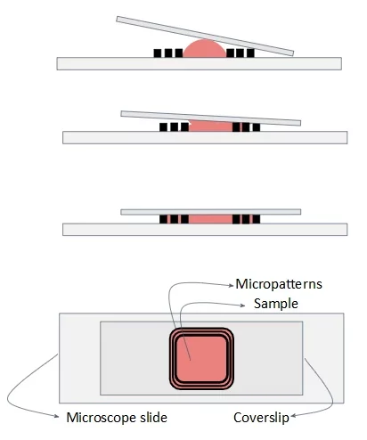
Micropatterns automatically remove air bubbles
Compatible with heat & enzyme treatments
Endures antigen retrieval procedures

Need a microfluidic SME partner for your Horizon Europe project?
Optimized microscope slides for high-quality immunohistochemistry
These trapped air pockets can hinder antibody penetration, disrupt the even distribution of reagents, and degrade image clarity in both immunohistochemistry and confocal microscopy, leading to unreliable results. Additionally, air pockets can cause inconsistent staining intensity, making quantitative analysis of biomarkers difficult and affecting diagnostic accuracy.
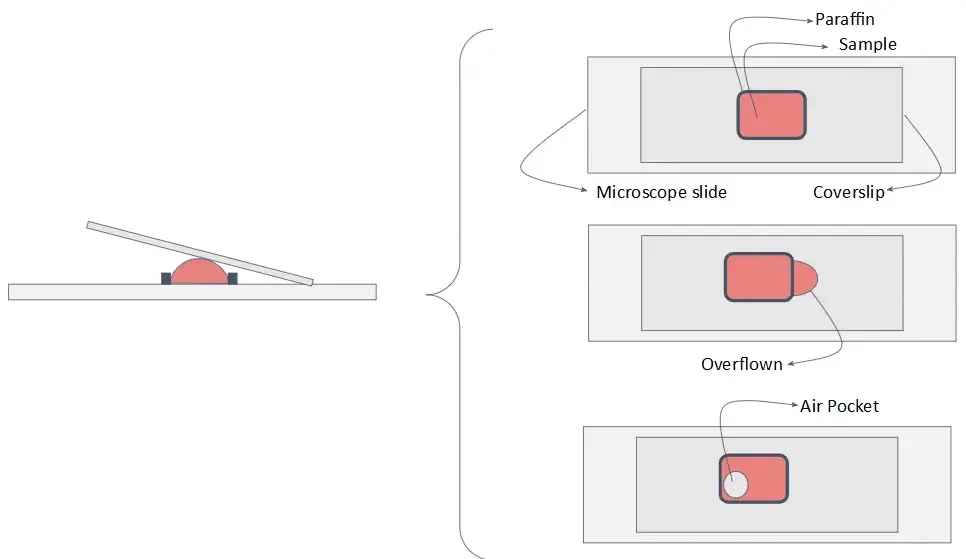
Additionally, capillary forces naturally hold the coverslip in place, eliminating the need for external sealants like wax or paraffin. This not only simplifies sample preparation but also preserves tissue morphology and enhances antibody interaction, leading to more precise and reproducible staining.
Moreover, the glass slide’s surface and micropattern design can be tailored to accommodate various sample types, making it an ideal solution for both standard and advanced Immunohistochemistry protocols.

References
Image credit: Detecting cancer in human tissues, LM. Aamir Ahmed, Jane Pendjiky and Michael Millar / Wellcome Collection
Compatibility and applications
Some other biological applications also include:
Histopathology
Clinical diagnostic
Immunofluorescence
Drug Screening
And many more!
Slides for immunohistochemistry protocols
The technical specifications of the product ensure air bubble-free imaging while maintaining high-quality staining and sample integrity for immunohistochemistry protocols.
| Technical Specifications | |
|---|---|
| Wetted materials | Glass or thermoplastics (PVD, CVD, PECVD) |
| Dimensions | Micropatterns:
|
| Working volumes | TBD |
Frequently asked questions
Can the working volume of the microscope slides be altered for different Immunohistochemistry protocols?
Yes, our slides are designed to accommodate different tissue section sizes and staining volumes. Contact our team for more information.
Do they work with any type of coverslip?
Yes, currently our slides are made on demand, so we can adjust the features to any coverslip dimensions. In any case, they are compatible with standard coverslips.
Can the shape of the micropatterns and observation chamber be altered?
Yes, contact our team for more information on custom designs.

Funding and Support
The ALTERNATIVE project developments helped develop this instrument. This project is funded by the European Union’s H2020-LC-GD-2020-3, grant agreement No. 101037090.
Products & Associated Accessories
FAQ - Slides for Immunohistochemistry Protocol
In simple words, what are these slides?
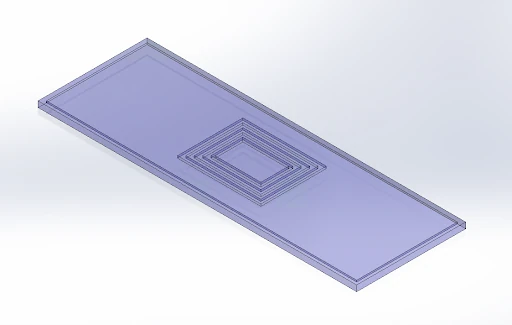
Micropatterned slides are glass sample holders with engraved microgrooves or patterns strategically placed around the sample viewing region. The rationale behind these slides is that they prevent air bubble entrapment during immunohistochemistry (IHC) and confocal microscopy, thereby providing high-quality staining and imaging of samples.
What would be so important about air bubbles in IHC and confocal imaging?
Since a trapped bubble can prevent antibody penetration in a local area, it will distort the distribution of reagents and produce patchy staining. Bubbles and patchy wetting also affect image quality and render quantitative comparisons (signal intensity, marker distribution) misleadingly unreliable in confocal microscopy.
Does it have multi-target staining (multiplex IHC)?
Yes, this is one of the major reasons. Even distribution of reagents and uniform wetting under the coverslip are just what you want when you are running sequential incubations or multi-target protocols, so the wetting between steps is the same.
Do I still need any external sealants, such as wax or paraffin?
Typically no. The design is based on capillary forces to hold the coverslip in place and retain liquid within the desired zone. The absence of the necessity of using wax/paraffin seals can help streamline workflows, speed them up, and (most importantly) reduce the error rate, particularly when you have to use an identical protocol on a large number of slides.
Do they work with the antigen retrieval (heat- and enzyme-based steps)?
Yes. The slides are said to be heat- and enzyme-compatible and designed to withstand antigen-retrieval conditions. This is important since ad-hoc chambers frequently fail by antigen retrieval (leaks, delamination, uneven wetting after heating, etc.).
What are the technical attributes of the micropatterns?
The following are the dimensions of the micropatterns listed:
- Depth: 100 um
- Width: 100 um
- Spacing of micropattern: 100 um
Wetted materials are denoted as glass or thermoplastics, and thin-film processes/coatings are compatible with PVD, CVD, and PECVD. The volumes are denoted as TBD (so volume is probably application- and geometry-dependent).
Which applications can these slides be used with?
The slides can be used with various biological applications:
- Immunohistochemistry (IHC)
- Multi-target IHC Multiplex staining (Multi-target IHC)
- Confocal microscopy
- Histopathology
- Clinical diagnostics
- Immunofluorescence
- Drug screening
- Antigen retrieval methods (heat and enzyme)
What are the major benefits of the micropatterned slides?
Bubble-free micropattern design: automatic removal.
- Streamlined operation: No external sealants are required.
- Improved and reproducible staining.
- Preservation: Maintains the tissue’s morphology.
- Increased communication: Better penetration by antibodies.
- Multiplex ready: Multi-target protocols.
- Versatility: Versatile to different types of samples.
Their work is with my coverslips, or do I have to use a particular format?
They are compatible with regular coverslips, and the slides are fabricated on request- that is to say, features can be scaled to suit the various sizes of coverslips. When you are in a lab where normal is not really normal (various thicknesses, unusually shaped, special-purpose optics), then this flexibility proves more useful than it may sound.
Is the chamber shape configurable to my tissue, sample type, or workflow?
Yes. The shape of the micropatterns and the observation chamber can be modified upon request. This is typically the distinction between a good idea and something you can actually use in a real-world consortium workflow (two or more partners, two or more sample types, two or more protocols).
What do I need to do before contacting MIC to get a quote or a personalized discussion about the product?
To have the quickest, most topical answer, come with:
- Size and thickness (usual range) of tissue section.
- Type of your coverslip (dimensions and thickness)
Do you heat retrieve, enzyme retrieve or both?
- Target range of working volume (it can be close to average)
- Imaging limitations (objective working distance, confocal vs widefield, immersion media).
